387D

RNA Pseudoknot with 3D Domain Swapping


Experimental Data Snapshot

  • Method: X-RAY DIFFRACTION
  • Resolution: 3.10 Å
  • R-Value Free: 0.234 
  • R-Value Work: 0.181 
  • R-Value Observed: 0.181 

wwPDB Validation   3D Report Full Report


This is version 1.4 of the entry. See complete history


Literature

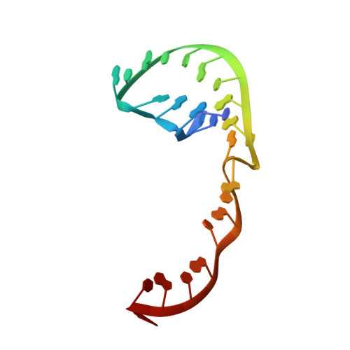
The Structure of an RNA Pseudoknot Shows 3D Domain Swapping

Lietzke, S.E.Barnes, C.L.Malone, V.F.Jones, J.T.Kundrot, C.E.

(1998) Structure, Motion, Interaction And Expression Of Biological Macromolecules, The Proceedings Of The Tenth Conversation Held At The University-suny, Albany Ny, June 17-21, 1997 10: 91-101


Macromolecules
Find similar nucleic acids by:  (by identity cutoff)  |  3D Structure
Entity ID: 1
MoleculeChains LengthOrganismImage
RNA Pseudoknot26N/A
Sequence Annotations
Expand
  • Reference Sequence
Experimental Data & Validation

Experimental Data

  • Method: X-RAY DIFFRACTION
  • Resolution: 3.10 Å
  • R-Value Free: 0.234 
  • R-Value Work: 0.181 
  • R-Value Observed: 0.181 
  • Space Group: P 43 2 2
Unit Cell:
Length ( Å )Angle ( ˚ )
a = 61.6α = 90
b = 61.6β = 90
c = 98.9γ = 90
Software Package:
Software NamePurpose
XDSdata scaling
XSCALEdata scaling
X-PLORrefinement
XDSdata reduction

Structure Validation

View Full Validation Report


Entry History 

Deposition Data

Revision History  (Full details and data files)

  • Version 1.0: 2003-08-26
    Type: Initial release
  • Version 1.1: 2008-04-26
    Changes: Version format compliance
  • Version 1.2: 2011-07-13
    Changes: Version format compliance
  • Version 1.3: 2018-04-04
    Changes: Data collection
  • Version 1.4: 2023-12-27
    Changes: Data collection, Database references