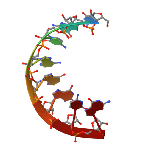
333D

THE CRYSTAL STRUCTURE OF AN RNA OLIGOMER INCORPORATING TANDEM ADENOSINE-INOSINE MISMATCHES


Experimental Data Snapshot

  • Method: X-RAY DIFFRACTION
  • Resolution: 2.52 Å
  • R-Value Free: 0.266 
  • R-Value Work: 0.246 
  • R-Value Observed: 0.246 

wwPDB Validation   3D Report Full Report


This is version 2.1 of the entry. See complete history


Literature

The crystal structure of an RNA oligomer incorporating tandem adenosine-inosine mismatches.

Carter, R.J.Baeyens, K.J.SantaLucia, J.Turner, D.H.Holbrook, S.R.

(1997) Nucleic Acids Res 25: 4117-4122

  • DOI: https://doi.org/10.1093/nar/25.20.4117
  • Primary Citation of Related Structures:  
    333D

  • PubMed Abstract: 

    The X-ray crystallographic structure of the RNA duplex [r(CGCAIGCG)]2 has been refined to 2.5 A. It shows a symmetric internal loop of two non-Watson-Crick base pairs which form in the middle of the duplex. The tandem A-I/I-A pairs are related by a crystallographic two-fold axis. Both A(anti)-I(anti) mismatches are in a head-to-head conformation forming hydrogen bonds using the Watson-Crick positions. The octamer duplexes stack above one another in the cell forming a pseudo-infinite helix throughout the crystal. A hydrated calcium ion bridges between the 3'-terminal of one molecule and the backbone of another. The tandem A-I mismatches are incorporated with only minor distortion to the backbone. This is in contrast to the large helical perturbations often produced by sheared G-A pairs in RNA oligonucleotides.


  • Organizational Affiliation

    Structural Biology Division, Lawrence Berkeley Laboratory, Berkeley, CA 94720, USA.


Macromolecules

Find similar nucleic acids by:  Sequence   |   3D Structure  

Entity ID: 1
MoleculeChains LengthOrganismImage
RNA (5'-R(*CP*GP*CP*AP*IP*GP*CP*G)-3')8N/A
Sequence Annotations
Expand
  • Reference Sequence
Small Molecules
Ligands 1 Unique
IDChains Name / Formula / InChI Key2D Diagram3D Interactions
CA
Query on CA

Download Ideal Coordinates CCD File 
B [auth A]CALCIUM ION
Ca
BHPQYMZQTOCNFJ-UHFFFAOYSA-N
Experimental Data & Validation

Experimental Data

  • Method: X-RAY DIFFRACTION
  • Resolution: 2.52 Å
  • R-Value Free: 0.266 
  • R-Value Work: 0.246 
  • R-Value Observed: 0.246 
  • Space Group: P 61 2 2
Unit Cell:
Length ( Å )Angle ( ˚ )
a = 39.05α = 90
b = 39.05β = 90
c = 58.85γ = 120
Software Package:
Software NamePurpose
X-PLORmodel building
X-PLORrefinement
R-AXISdata reduction
X-PLORphasing

Structure Validation

View Full Validation Report


Entry History 

Deposition Data

Revision History  (Full details and data files)

  • Version 1.0: 1997-10-16
    Type: Initial release
  • Version 1.1: 2008-05-22
    Changes: Version format compliance
  • Version 1.2: 2011-07-13
    Changes: Version format compliance
  • Version 2.0: 2024-02-21
    Changes: Atomic model, Data collection, Database references, Derived calculations
  • Version 2.1: 2024-04-03
    Changes: Refinement description