2ZZL

Structure of bacteriorhodopsin's M intermediate at pH 7


Experimental Data Snapshot

  • Method: X-RAY DIFFRACTION
  • Resolution: 2.03 Å
  • R-Value Free: 0.238 
  • R-Value Work: 0.207 
  • R-Value Observed: 0.207 

wwPDB Validation   3D Report Full Report


This is version 2.1 of the entry. See complete history


Literature

Crystal Structures of Different Substates of Bacteriorhodopsin's M Intermediate at Various pH Levels.

Yamamoto, M.Hayakawa, N.Murakami, M.Kouyama, T.

(2009) J Mol Biol 

  • DOI: https://doi.org/10.1016/j.jmb.2009.08.047
  • Primary Citation of Related Structures:  
    2ZZL

  • PubMed Abstract: 

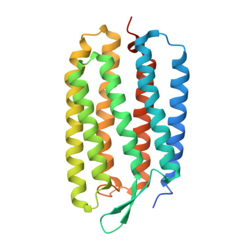
    The hexagonal P622 crystal of bacteriorhodopsin, which is made up of stacked membranes, is stable provided that the precipitant concentration in the soaking solution is higher than a critical value (i.e., 1.5 M ammonium sulfate). Diffraction data showed that the crystal lattice shrank linearly with increasing precipitant concentration, due primarily to narrowing of intermembrane spaces. Although the crystal shrinkage did not affect the rate of formation of the photoreaction M intermediate, its lifetime increased exponentially with the precipitant concentration. It was suggested that the energetic barrier of the M-to-N transition becomes higher when the motional freedom of the EF loop is reduced by crystal lattice force. As a result of this property, the M state accumulated predominantly when the crystal that was soaked at a high precipitant concentration was illuminated at room temperature. Structural data obtained at various pH levels showed that the overall structure of M is not strongly dependent on pH, except that Glu194 and Glu204 in the proton release complex are more separated at pH 7 than at pH 4.4. This result suggests that light-induced disruption of the paired structure of Glu194 and Glu204 is incomplete when external pH is lower than the pK(a) value of the proton release group in the M state.


  • Organizational Affiliation

    Department of Physics, Graduate School of Science, Nagoya University, Nagoya, Japan.


Macromolecules
Find similar proteins by:  (by identity cutoff)  |  3D Structure
Entity ID: 1
MoleculeChains Sequence LengthOrganismDetailsImage
Bacteriorhodopsin262Halobacterium salinarumMutation(s): 0 
Membrane Entity: Yes 
UniProt
Find proteins for P02945 (Halobacterium salinarum (strain ATCC 700922 / JCM 11081 / NRC-1))
Explore P02945 
Go to UniProtKB:  P02945
Entity Groups  
Sequence Clusters30% Identity50% Identity70% Identity90% Identity95% Identity100% Identity
UniProt GroupP02945
Sequence Annotations
Expand
  • Reference Sequence
Oligosaccharides

Help

Entity ID: 2
MoleculeChains Length2D Diagram Glycosylation3D Interactions
3-O-sulfo-beta-D-galactopyranose-(1-6)-alpha-D-mannopyranose-(1-2)-alpha-D-glucopyranose
B
3N/A
Glycosylation Resources
GlyTouCan:  G37414ZM
GlyCosmos:  G37414ZM
Small Molecules
Ligands 5 Unique
IDChains Name / Formula / InChI Key2D Diagram3D Interactions
L3P
Query on L3P

Download Ideal Coordinates CCD File 
D [auth A]2,3-DI-O-PHYTANLY-3-SN-GLYCERO-1-PHOSPHORYL-3'-SN-GLYCEROL-1'-PHOSPHATE
C46 H94 O11 P2
TZXJQSKPTCRGCA-VZSPAKCESA-L
L1P
Query on L1P

Download Ideal Coordinates CCD File 
F [auth A],
G [auth A],
H [auth A]
3-PHOSPHORYL-[1,2-DI-PHYTANYL]GLYCEROL
C43 H89 O6 P
UKQGAMWGTOTQPC-ALOLAALWSA-N
L2P
Query on L2P

Download Ideal Coordinates CCD File 
E [auth A]2,3-DI-PHYTANYL-GLYCEROL
C43 H88 O3
ISDBCJSGCHUHFI-UMZPFTBHSA-N
SOG
Query on SOG

Download Ideal Coordinates CCD File 
I [auth A]octyl 1-thio-beta-D-glucopyranoside
C14 H28 O5 S
CGVLVOOFCGWBCS-RGDJUOJXSA-N
RET
Query on RET

Download Ideal Coordinates CCD File 
C [auth A]RETINAL
C20 H28 O
NCYCYZXNIZJOKI-OVSJKPMPSA-N
Experimental Data & Validation

Experimental Data

  • Method: X-RAY DIFFRACTION
  • Resolution: 2.03 Å
  • R-Value Free: 0.238 
  • R-Value Work: 0.207 
  • R-Value Observed: 0.207 
  • Space Group: P 6 2 2
Unit Cell:
Length ( Å )Angle ( ˚ )
a = 102.21α = 90
b = 102.21β = 90
c = 112.48γ = 120
Software Package:
Software NamePurpose
MAR345dtbdata collection
CNSrefinement
MOSFLMdata reduction
SCALAdata scaling
CNSphasing

Structure Validation

View Full Validation Report


Entry History 

Deposition Data

Revision History  (Full details and data files)

  • Version 1.0: 2009-10-06
    Type: Initial release
  • Version 1.1: 2011-07-13
    Changes: Version format compliance
  • Version 2.0: 2020-07-29
    Type: Remediation
    Reason: Carbohydrate remediation
    Changes: Advisory, Atomic model, Data collection, Derived calculations, Structure summary
  • Version 2.1: 2023-11-01
    Changes: Data collection, Database references, Refinement description, Structure summary