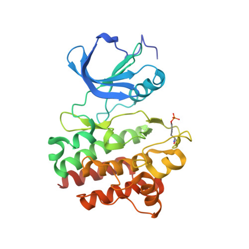
2ZYB

Crystal structure of phenylimidazo pyrazin 2 bound to the kinase domain of human LCK, (auto-phosphorylated on TYR394)


Experimental Data Snapshot

  • Method: X-RAY DIFFRACTION
  • Resolution: 2.55 Å
  • R-Value Free: 0.291 
  • R-Value Work: 0.171 
  • R-Value Observed: 0.183 

wwPDB Validation   3D Report Full Report


Ligand Structure Quality Assessment 


This is version 1.3 of the entry. See complete history


Literature

The importance of CH/pi hydrogen bonds in rational drug design: An ab initio fragment molecular orbital study to leukocyte-specific protein tyrosine (LCK) kinase

Ozawa, T.Tsuji, E.Ozawa, M.Handa, C.Mukaiyama, H.Nishimura, T.Kobayashi, S.Okazaki, K.

(2008) Bioorg Med Chem 16: 10311-10318

  • DOI: https://doi.org/10.1016/j.bmc.2008.10.041
  • Primary Citation of Related Structures:  
    2ZM1, 2ZM4, 2ZYB

  • PubMed Abstract: 

    The interaction energy was calculated, by the ab initio FMO method, for complexes between LCK protein and four inhibitors (staurosporine, BMS compound 2, and our compounds 3 and 4). In every case a number of CH/pi hydrogen bonds have been disclosed in the so-called adenine pocket. In complexes of 2, 3, and 4, CH/pi and NH/pi hydrogen bonds have been observed in another pocket. In view of the above results, the aniline ring of 3 was replaced by 2,6-dimethyl aniline to increase the potency for LCK kinase. A 10-fold increase in the potency has been achieved for 4 over 3. We suggest that the concept of weak hydrogen bonds is useful in the rational design of drugs.


  • Organizational Affiliation

    Kissei Pharmaceutical Company Ltd., Central Research Laboratory, 4365-1 Kashiwabara, Hotaka, Azumino, Nagano-Pref 399-8304, Japan. tomonaga_ozawa@pharm.kissei.co.jp


Macromolecules
Find similar proteins by:  (by identity cutoff)  |  3D Structure
Entity ID: 1
MoleculeChains Sequence LengthOrganismDetailsImage
Proto-oncogene tyrosine-protein kinase LCK285Homo sapiensMutation(s): 0 
Gene Names: LCK
EC: 2.7.10.2
UniProt & NIH Common Fund Data Resources
Find proteins for P06239 (Homo sapiens)
Explore P06239 
Go to UniProtKB:  P06239
PHAROS:  P06239
GTEx:  ENSG00000182866 
Entity Groups  
Sequence Clusters30% Identity50% Identity70% Identity90% Identity95% Identity100% Identity
UniProt GroupP06239
Sequence Annotations
Expand
  • Reference Sequence
Small Molecules
Ligands 2 Unique
IDChains Name / Formula / InChI Key2D Diagram3D Interactions
KSL
Query on KSL

Download Ideal Coordinates CCD File 
C [auth A]N-(2,6-dimethylphenyl)-5-phenylimidazo[1,5-a]pyrazin-8-amine
C20 H18 N4
KKYYLKPGILUPOA-UHFFFAOYSA-N
SO4
Query on SO4

Download Ideal Coordinates CCD File 
B [auth A]SULFATE ION
O4 S
QAOWNCQODCNURD-UHFFFAOYSA-L
Modified Residues  1 Unique
IDChains TypeFormula2D DiagramParent
PTR
Query on PTR
A
L-PEPTIDE LINKINGC9 H12 N O6 PTYR
Binding Affinity Annotations 
IDSourceBinding Affinity
PTR BindingDB:  2ZYB Kd: 4.00e+5 (nM) from 1 assay(s)
KSL PDBBind:  2ZYB IC50: 20 (nM) from 1 assay(s)
BindingDB:  2ZYB IC50: 20 (nM) from 1 assay(s)
Experimental Data & Validation

Experimental Data

  • Method: X-RAY DIFFRACTION
  • Resolution: 2.55 Å
  • R-Value Free: 0.291 
  • R-Value Work: 0.171 
  • R-Value Observed: 0.183 
  • Space Group: P 21 21 21
Unit Cell:
Length ( Å )Angle ( ˚ )
a = 42.15α = 90
b = 73.973β = 90
c = 92.857γ = 90
Software Package:
Software NamePurpose
REFMACrefinement
BSSdata collection
MOSFLMdata reduction
SCALAdata scaling
AMoREphasing

Structure Validation

View Full Validation Report


Ligand Structure Quality Assessment 


Entry History 

Deposition Data

  • Released Date: 2009-02-03 
  • Deposition Author(s): Tsuji, E.

Revision History  (Full details and data files)

  • Version 1.0: 2009-02-03
    Type: Initial release
  • Version 1.1: 2011-07-13
    Changes: Version format compliance
  • Version 1.2: 2023-11-01
    Changes: Data collection, Database references, Derived calculations, Refinement description
  • Version 1.3: 2023-11-15
    Changes: Data collection