2ZY2

dodecameric L-aspartate beta-decarboxylase


Experimental Data Snapshot

  • Method: X-RAY DIFFRACTION
  • Resolution: 3.30 Å
  • R-Value Free: 0.293 
  • R-Value Work: 0.228 
  • R-Value Observed: 0.231 

wwPDB Validation   3D Report Full Report


Ligand Structure Quality Assessment 


This is version 1.2 of the entry. See complete history


Literature

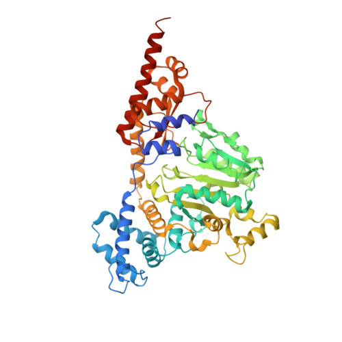
Structure, Assembly, and Mechanism of a PLP-Dependent Dodecameric l-Aspartate beta-Decarboxylase

Chen, H.-J.Ko, T.-P.Lee, C.-Y.Wang, N.-C.Wang, A.H.-J.

(2009) Structure 17: 517-529

  • DOI: https://doi.org/10.1016/j.str.2009.02.013
  • Primary Citation of Related Structures:  
    2ZY2, 2ZY3, 2ZY4, 2ZY5

  • PubMed Abstract: 

    The type-I PLP enzyme l-aspartate beta-decarboxylase converts aspartate to alanine and CO(2). Similar to the homodimeric aminotransferases, its protein subunit comprises a large and a small domain, of 410 and 120 residues, respectively. The crystal structure reveals a dodecamer made of six identical dimers arranged in a truncated tetrahedron whose assembly involves tetramer and hexamer as intermediates. The additional helical motifs I and II participate in the oligomer formation. Triple mutations of S67R/Y68R/M69R or S67E/Y68E/M69E in motif I produced an inactive dimer. The PLP is bound covalently to Lys315 in the active site, while its phosphate group interacts with a neighboring Tyr134. Removal of the bulky side chain of Arg37, which overhangs the PLP group, improved the substrate affinity. Mutations in flexible regions produced the more active K17A and the completely inactive R487A. The structure also suggests that substrate binding triggers conformational changes essential for catalyzing the reaction.


  • Organizational Affiliation

    Institute of Biological Chemistry, Academia Sinica, Taipei 11529, Taiwan.


Macromolecules
Find similar proteins by:  (by identity cutoff)  |  3D Structure
Entity ID: 1
MoleculeChains Sequence LengthOrganismDetailsImage
L-aspartate 4-carboxylyase544Pseudomonas sp. ATCC 19121Mutation(s): 0 
Gene Names: asdP
EC: 4.1.1.12
UniProt
Find proteins for Q53IZ1 (Pseudomonas sp)
Explore Q53IZ1 
Go to UniProtKB:  Q53IZ1
Entity Groups  
Sequence Clusters30% Identity50% Identity70% Identity90% Identity95% Identity100% Identity
UniProt GroupQ53IZ1
Sequence Annotations
Expand
  • Reference Sequence
Small Molecules
Ligands 1 Unique
IDChains Name / Formula / InChI Key2D Diagram3D Interactions
PLP
Query on PLP

Download Ideal Coordinates CCD File 
B [auth A]PYRIDOXAL-5'-PHOSPHATE
C8 H10 N O6 P
NGVDGCNFYWLIFO-UHFFFAOYSA-N
Experimental Data & Validation

Experimental Data

  • Method: X-RAY DIFFRACTION
  • Resolution: 3.30 Å
  • R-Value Free: 0.293 
  • R-Value Work: 0.228 
  • R-Value Observed: 0.231 
  • Space Group: F 4 3 2
Unit Cell:
Length ( Å )Angle ( ˚ )
a = 298.9α = 90
b = 298.9β = 90
c = 298.9γ = 90
Software Package:
Software NamePurpose
Blu-Icedata collection
SOLVEphasing
CNSrefinement
HKL-2000data reduction
HKL-2000data scaling

Structure Validation

View Full Validation Report


Ligand Structure Quality Assessment 


Entry History 

Deposition Data

Revision History  (Full details and data files)

  • Version 1.0: 2009-01-27
    Type: Initial release
  • Version 1.1: 2011-07-13
    Changes: Source and taxonomy, Version format compliance
  • Version 1.2: 2017-10-11
    Changes: Refinement description