2ZY1

Crystal structure of the C(30) carotenoid dehydrosqualene synthase from Staphylococcus aureus complexed with bisphosphonate BPH-830


Experimental Data Snapshot

  • Method: X-RAY DIFFRACTION
  • Resolution: 1.78 Å
  • R-Value Free: 0.219 
  • R-Value Work: 0.182 

wwPDB Validation   3D Report Full Report


Ligand Structure Quality Assessment 


This is version 1.2 of the entry. See complete history


Literature

Inhibition of staphyloxanthin virulence factor biosynthesis in Staphylococcus aureus: in vitro, in vivo, and crystallographic results.

Song, Y.Liu, C.I.Lin, F.Y.No, J.H.Hensler, M.Liu, Y.L.Jeng, W.Y.Low, J.Liu, G.Y.Nizet, V.Wang, A.H.J.Oldfield, E.

(2009) J Med Chem 52: 3869-3880

  • DOI: https://doi.org/10.1021/jm9001764
  • Primary Citation of Related Structures:  
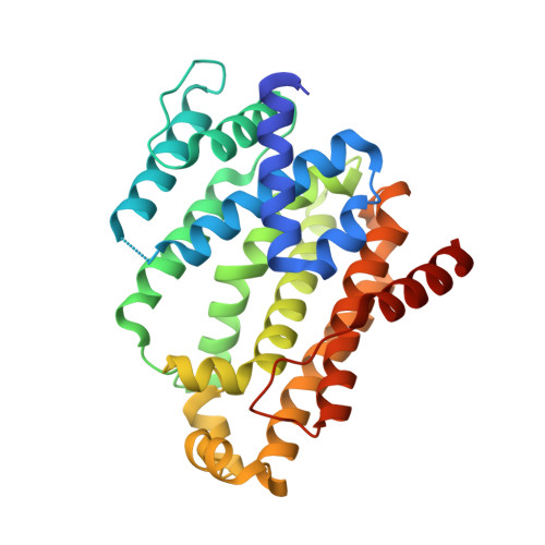
    2ZY1

  • PubMed Abstract: 

    The gold color of Staphylococcus aureus is derived from the carotenoid staphyloxanthin, a virulence factor for the organism. Here, we report the synthesis and activity of a broad variety of staphyloxanthin biosynthesis inhibitors that inhibit the first committed step in its biosynthesis, condensation of two farnesyl diphosphate (FPP) molecules to dehydrosqualene, catalyzed by the enzyme dehydrosqualene synthase (CrtM). The most active compounds are phosphonoacetamides that have low nanomolar K(i) values for CrtM inhibition and are active in whole bacterial cells and in mice, where they inhibit S. aureus disease progression. We also report the X-ray crystallographic structure of the most active compound, N-3-(3-phenoxyphenyl)propylphosphonoacetamide (IC(50) = 8 nM, in cells), bound to CrtM. The structure exhibits a complex network of hydrogen bonds between the polar headgroup and the protein, while the 3-phenoxyphenyl side chain is located in a hydrophobic pocket previously reported to bind farnesyl thiodiphosphate (FsPP), as well as biphenyl phosphonosulfonate inhibitors. Given the good enzymatic, whole cell, and in vivo pharmacologic activities, these results should help guide the further development of novel antivirulence factor-based therapies for S. aureus infections.


  • Organizational Affiliation

    Department of Chemistry and Center for Biophysics and Computational Biology, University of Illinois at Urbana-Champaign, Urbana, Illinois 61801, USA.


Macromolecules
Find similar proteins by:  (by identity cutoff)  |  3D Structure
Entity ID: 1
MoleculeChains Sequence LengthOrganismDetailsImage
Dehydrosqualene synthase293Staphylococcus aureusMutation(s): 0 
Gene Names: crtM
EC: 2.5.1
UniProt
Find proteins for A9JQL9 (Staphylococcus aureus)
Explore A9JQL9 
Go to UniProtKB:  A9JQL9
Entity Groups  
Sequence Clusters30% Identity50% Identity70% Identity90% Identity95% Identity100% Identity
UniProt GroupA9JQL9
Sequence Annotations
Expand
  • Reference Sequence
Small Molecules
Ligands 1 Unique
IDChains Name / Formula / InChI Key2D Diagram3D Interactions
830
Query on 830

Download Ideal Coordinates CCD File 
B [auth A]dipotassium (2-oxo-2-{[3-(3-phenoxyphenyl)propyl]amino}ethyl)phosphonate
C17 H18 K2 N O5 P
VIIVNJCQPLUIGT-UHFFFAOYSA-L
Binding Affinity Annotations 
IDSourceBinding Affinity
830 Binding MOAD:  2ZY1 Ki: 40 (nM) from 1 assay(s)
PDBBind:  2ZY1 Ki: 40 (nM) from 1 assay(s)
Experimental Data & Validation

Experimental Data

  • Method: X-RAY DIFFRACTION
  • Resolution: 1.78 Å
  • R-Value Free: 0.219 
  • R-Value Work: 0.182 
  • Space Group: P 32 2 1
Unit Cell:
Length ( Å )Angle ( ˚ )
a = 80.59α = 90
b = 80.59β = 90
c = 90.02γ = 120
Software Package:
Software NamePurpose
HKL-2000data collection
CNSrefinement
HKL-2000data reduction
HKL-2000data scaling
CNSphasing

Structure Validation

View Full Validation Report


Ligand Structure Quality Assessment 


Entry History 

Deposition Data

Revision History  (Full details and data files)

  • Version 1.0: 2009-09-01
    Type: Initial release
  • Version 1.1: 2011-07-13
    Changes: Version format compliance
  • Version 1.2: 2023-11-01
    Changes: Data collection, Database references, Derived calculations, Refinement description