


Novel Antibiotics Target Protein of Gram-positive Pathogens: X-ray Crystal Structures and Search of Potential Drug-Binding Sites
Doi, A., Okajima, T., Gotoh, Y., Tanizawa, K., Utsumi, R.To be published.
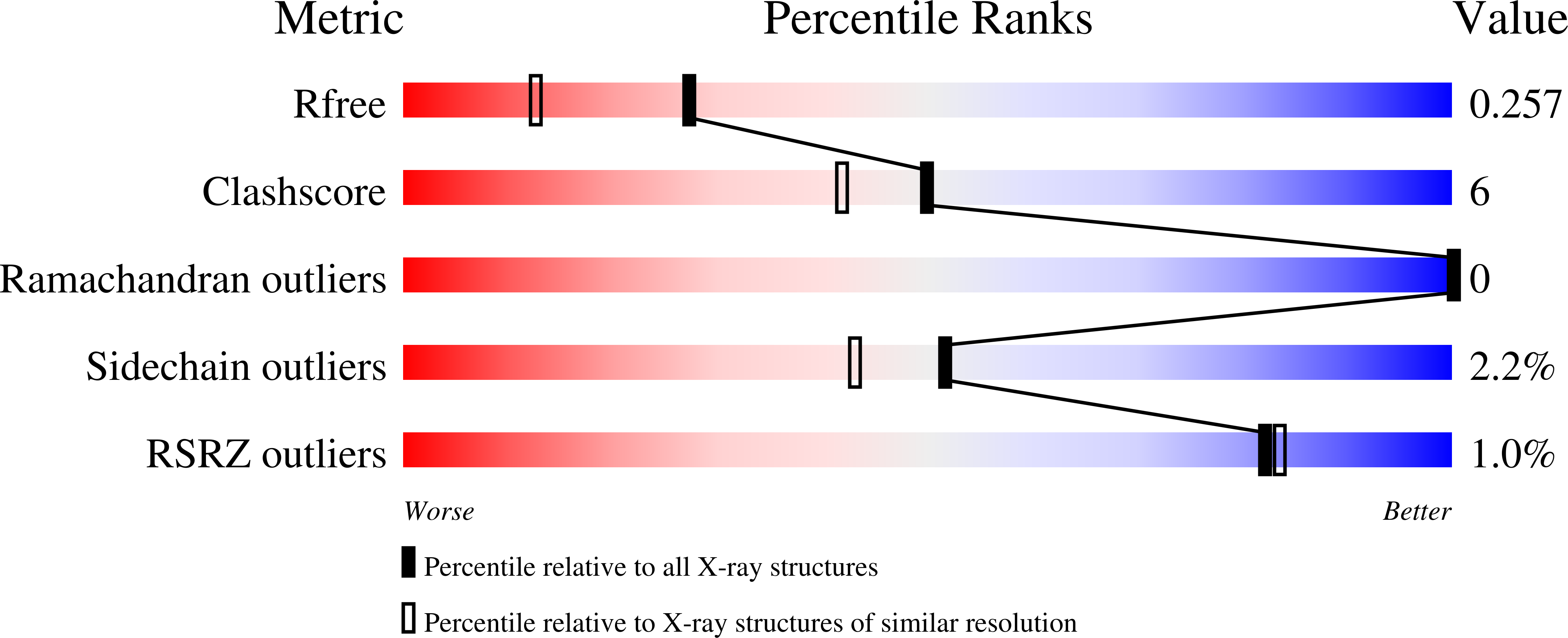
Experimental Data Snapshot
wwPDB Validation 3D Report Full Report
Entity ID: 1 | |||||
|---|---|---|---|---|---|
| Molecule | Chains | Sequence Length | Organism | Details | Image |
| Transcriptional regulatory protein walR | 120 | Staphylococcus aureus | Mutation(s): 0 Gene Names: yycF |  | |
UniProt | |||||
Find proteins for Q9RDT5 (Staphylococcus aureus) Explore Q9RDT5 Go to UniProtKB: Q9RDT5 | |||||
Entity Groups | |||||
| Sequence Clusters | 30% Identity50% Identity70% Identity90% Identity95% Identity100% Identity | ||||
| UniProt Group | Q9RDT5 | ||||
Sequence AnnotationsExpand | |||||
| |||||
| Length ( Å ) | Angle ( ˚ ) |
|---|---|
| a = 65.634 | α = 90 |
| b = 45.556 | β = 103.52 |
| c = 78.043 | γ = 90 |
| Software Name | Purpose |
|---|---|
| REFMAC | refinement |
| MOSFLM | data reduction |
| SCALA | data scaling |
| PHASER | phasing |
RCSB PDB (citation) is hosted by
RCSB PDB is a member of the

