


Novel Antibiotics Target Protein of Gram-positive Pathogens: X-ray Crystal Structures and Search of Potential Drug-Binding sites
Doi, A., Okajima, T., Gotoh, Y., Tanizawa, K., Utsumi, R.To be published.
Experimental Data Snapshot
wwPDB Validation 3D Report Full Report

Entity ID: 1 | |||||
|---|---|---|---|---|---|
| Molecule | Chains | Sequence Length | Organism | Details | Image |
| Transcriptional regulatory protein yycF | 130 | Bacillus subtilis | Mutation(s): 0 Gene Names: yycF | 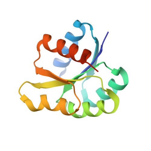 | |
UniProt | |||||
Find proteins for P37478 (Bacillus subtilis (strain 168)) Explore P37478 Go to UniProtKB: P37478 | |||||
Entity Groups | |||||
| Sequence Clusters | 30% Identity50% Identity70% Identity90% Identity95% Identity100% Identity | ||||
| UniProt Group | P37478 | ||||
Sequence AnnotationsExpand | |||||
| |||||
| Ligands 1 Unique | |||||
|---|---|---|---|---|---|
| ID | Chains | Name / Formula / InChI Key | 2D Diagram | 3D Interactions | |
| SO4 Query on SO4 | C [auth A], D [auth A], E [auth B], F [auth B] | SULFATE ION O4 S QAOWNCQODCNURD-UHFFFAOYSA-L |  | ||
| Length ( Å ) | Angle ( ˚ ) |
|---|---|
| a = 58.824 | α = 90 |
| b = 58.824 | β = 90 |
| c = 78.247 | γ = 120 |
| Software Name | Purpose |
|---|---|
| REFMAC | refinement |
| MOSFLM | data reduction |
| SCALA | data scaling |
| PHASER | phasing |
RCSB PDB (citation) is hosted by
RCSB PDB is a member of the

