2ZWH

Model for the F-actin structure


Experimental Data Snapshot

  • Method: FIBER DIFFRACTION
  • Resolution: 3.30 Å

wwPDB Validation   3D Report Full Report


This is version 1.4 of the entry. See complete history


Literature

The nature of the globular- to fibrous-actin transition.

Oda, T.Iwasa, M.Aihara, T.Maeda, Y.Narita, A.

(2009) Nature 457: 441-445

  • DOI: https://doi.org/10.1038/nature07685
  • Primary Citation of Related Structures:  
    2ZWH

  • PubMed Abstract: 

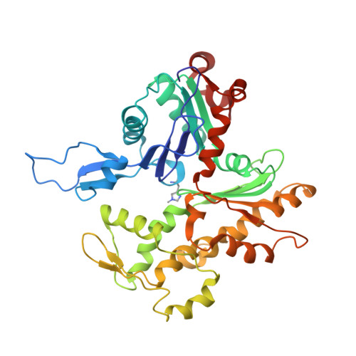
    Actin plays crucial parts in cell motility through a dynamic process driven by polymerization and depolymerization, that is, the globular (G) to fibrous (F) actin transition. Although our knowledge about the actin-based cellular functions and the molecules that regulate the G- to F-actin transition is growing, the structural aspects of the transition remain enigmatic. We created a model of F-actin using X-ray fibre diffraction intensities obtained from well oriented sols of rabbit skeletal muscle F-actin to 3.3 A in the radial direction and 5.6 A along the equator. Here we show that the G- to F-actin conformational transition is a simple relative rotation of the two major domains by about 20 degrees. As a result of the domain rotation, the actin molecule in the filament is flat. The flat form is essential for the formation of stable, helical F-actin. Our F-actin structure model provides the basis for understanding actin polymerization as well as its molecular interactions with actin-binding proteins.


  • Organizational Affiliation

    X-ray Structural Analysis Research Team, RIKEN SPring-8 Center, RIKEN Harima Institute, 1-1-1, Kouto, Sayo, Hyogo 679-5148, Japan. toda@spring8.or.jp


Macromolecules
Find similar proteins by:  (by identity cutoff)  |  3D Structure
Entity ID: 1
MoleculeChains Sequence LengthOrganismDetailsImage
Actin, alpha skeletal muscle375Oryctolagus cuniculusMutation(s): 0 
UniProt
Find proteins for P68135 (Oryctolagus cuniculus)
Explore P68135 
Go to UniProtKB:  P68135
Entity Groups  
Sequence Clusters30% Identity50% Identity70% Identity90% Identity95% Identity100% Identity
UniProt GroupP68135
Sequence Annotations
Expand
  • Reference Sequence
Small Molecules
Ligands 2 Unique
IDChains Name / Formula / InChI Key2D Diagram3D Interactions
ADP
Query on ADP

Download Ideal Coordinates CCD File 
B [auth A]ADENOSINE-5'-DIPHOSPHATE
C10 H15 N5 O10 P2
XTWYTFMLZFPYCI-KQYNXXCUSA-N
CA
Query on CA

Download Ideal Coordinates CCD File 
C [auth A]CALCIUM ION
Ca
BHPQYMZQTOCNFJ-UHFFFAOYSA-N
Modified Residues  1 Unique
IDChains TypeFormula2D DiagramParent
HIC
Query on HIC
A
L-PEPTIDE LINKINGC7 H11 N3 O2HIS
Experimental Data & Validation

Experimental Data

  • Method: FIBER DIFFRACTION
  • Resolution: 3.30 Å
  • Space Group: P 1
Unit Cell:
Length ( Å )Angle ( ˚ )
a = 1α = 90
b = 1β = 90
c = 1γ = 90
Software Package:
Software NamePurpose
X-PLORrefinement

Structure Validation

View Full Validation Report


Entry History 

Deposition Data

Revision History  (Full details and data files)

  • Version 1.0: 2009-01-20
    Type: Initial release
  • Version 1.1: 2011-07-13
    Changes: Version format compliance
  • Version 1.2: 2015-02-11
    Changes: Other
  • Version 1.3: 2015-10-14
    Changes: Refinement description
  • Version 1.4: 2017-10-11
    Changes: Refinement description