2ZTL

Closed conformation of D-3-hydroxybutyrate dehydrogenase complexed with NAD+ and L-3-hydroxybutyrate


Experimental Data Snapshot

  • Method: X-RAY DIFFRACTION
  • Resolution: 1.80 Å
  • R-Value Free: 0.229 
  • R-Value Work: 0.198 
  • R-Value Observed: 0.200 

wwPDB Validation   3D Report Full Report


Ligand Structure Quality Assessment 


This is version 1.2 of the entry. See complete history


Literature

Closed complex of the D-3-hydroxybutyrate dehydrogenase induced by an enantiomeric competitive inhibitor.

Nakashima, K.Ito, K.Nakajima, Y.Yamazawa, R.Miyakawa, S.Yoshimoto, T.

(2009) J Biochem 145: 467-479

  • DOI: https://doi.org/10.1093/jb/mvn186
  • Primary Citation of Related Structures:  
    2ZTL, 2ZTM, 2ZTU, 2ZTV

  • PubMed Abstract: 

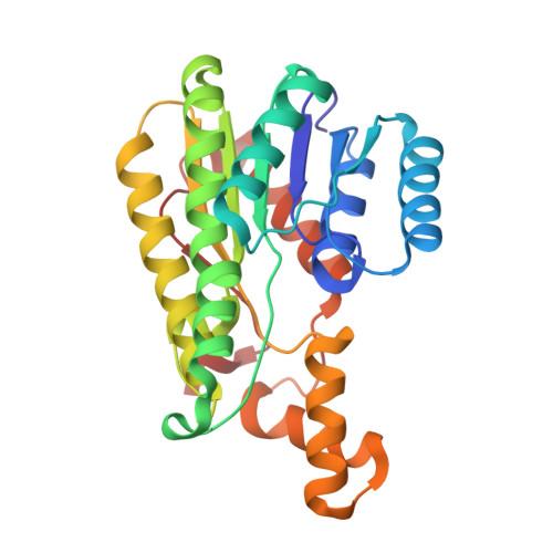
    D-3-Hydroxybutyrate dehydrogenase (HBDH) from Pseudomonas fragi showed a strict stereospecificity to the d-enantiomer of 3-hydroxybutyrate (d-3-HB) as a substrate. The l-enantiomer acts as a competitive inhibitor, with a K(i) value comparable to the K(m) value for d-3-HB. We have determined the crystal structures of the ternary complex of HBDH-NAD(+)-l-3-HB and the binary complex of HBDH-NAD(+). The former structure showed a so-called closed-form conformation, which is considered an active form for catalysis, while the latter stayed mostly in a open-form conformation. The determined structures along with the site-directed mutagenesis confirmed the substrate recognition mechanism that we proposed previously. The hydrogen bonding interaction between Gln196, located in the moving helix, and the carboxyl group of the substrate/inhibitor is important for the stable ternary complex formation. Finally, the crystal structures of the Thr190 mutants, T190S and T190A, indicate that the Thr190 is a key residue for the open-closed conformational change. T190S retained 37% of the activity. In T190A, however, the activity decreased to 0.1% that of the wild-type enzyme. Fixing the position of the hydroxyl group of Thr190 to form hydrogen bonds to the pyrophosphate moiety and the carboxamide of NAD(+) seems to be a significant factor for the open-closed conformational change.


  • Organizational Affiliation

    Department of Molecular Medicinal Sciences, Graduate School of Biomedical Sciences, Nagasaki University, 1-14 Bunkyo-machi, Nagasaki 852-8521, Japan.


Macromolecules
Find similar proteins by:  (by identity cutoff)  |  3D Structure
Entity ID: 1
MoleculeChains Sequence LengthOrganismDetailsImage
D(-)-3-hydroxybutyrate dehydrogenase
A, B, C, D
260Pseudomonas fragiMutation(s): 0 
EC: 1.1.1.30
UniProt
Find proteins for Q5KST5 (Pseudomonas fragi)
Explore Q5KST5 
Go to UniProtKB:  Q5KST5
Entity Groups  
Sequence Clusters30% Identity50% Identity70% Identity90% Identity95% Identity100% Identity
UniProt GroupQ5KST5
Sequence Annotations
Expand
  • Reference Sequence
Small Molecules
Ligands 4 Unique
IDChains Name / Formula / InChI Key2D Diagram3D Interactions
NAD
Query on NAD

Download Ideal Coordinates CCD File 
E [auth A],
H [auth B],
K [auth C],
M [auth D]
NICOTINAMIDE-ADENINE-DINUCLEOTIDE
C21 H27 N7 O14 P2
BAWFJGJZGIEFAR-NNYOXOHSSA-N
3HL
Query on 3HL

Download Ideal Coordinates CCD File 
F [auth A],
I [auth B],
L [auth C],
N [auth D]
(3S)-3-HYDROXYBUTANOIC ACID
C4 H8 O3
WHBMMWSBFZVSSR-VKHMYHEASA-N
GOL
Query on GOL

Download Ideal Coordinates CCD File 
O [auth D]GLYCEROL
C3 H8 O3
PEDCQBHIVMGVHV-UHFFFAOYSA-N
MG
Query on MG

Download Ideal Coordinates CCD File 
G [auth A],
J [auth B]
MAGNESIUM ION
Mg
JLVVSXFLKOJNIY-UHFFFAOYSA-N
Experimental Data & Validation

Experimental Data

  • Method: X-RAY DIFFRACTION
  • Resolution: 1.80 Å
  • R-Value Free: 0.229 
  • R-Value Work: 0.198 
  • R-Value Observed: 0.200 
  • Space Group: P 21 21 21
Unit Cell:
Length ( Å )Angle ( ˚ )
a = 74.018α = 90
b = 106.384β = 90
c = 165.168γ = 90
Software Package:
Software NamePurpose
REFMACrefinement
ADSCdata collection
HKL-2000data reduction
HKL-2000data scaling
MOLREPphasing

Structure Validation

View Full Validation Report


Ligand Structure Quality Assessment 


Entry History 

Deposition Data

Revision History  (Full details and data files)

  • Version 1.0: 2009-08-25
    Type: Initial release
  • Version 1.1: 2011-07-13
    Changes: Non-polymer description, Version format compliance
  • Version 1.2: 2023-11-01
    Changes: Data collection, Database references, Derived calculations, Refinement description