2ZTH

Crystal structure of holo form of rat catechol-o-methyltransferase


Experimental Data Snapshot

  • Method: X-RAY DIFFRACTION
  • Resolution: 2.60 Å
  • R-Value Free: 0.281 
  • R-Value Work: 0.187 
  • R-Value Observed: 0.196 

wwPDB Validation   3D Report Full Report


Ligand Structure Quality Assessment 


This is version 1.2 of the entry. See complete history


Literature

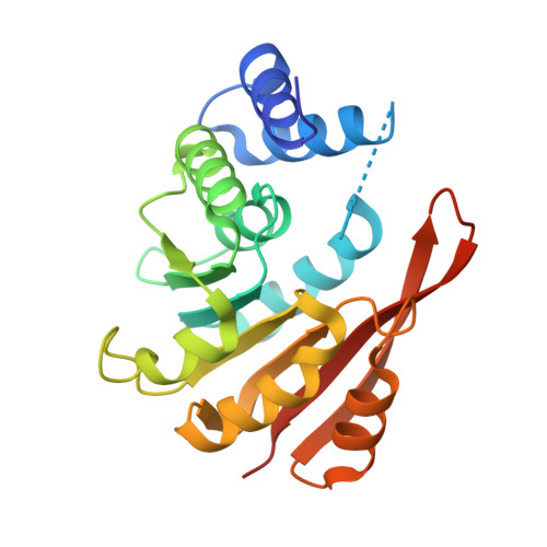
Crystal structures of the Apo and Holo form of rat catechol-O-methyltransferase

Tsuji, E.Okazaki, K.Isaji, M.Takeda, K.

(2009) J Struct Biol 165: 133-139

  • DOI: https://doi.org/10.1016/j.jsb.2008.11.012
  • Primary Citation of Related Structures:  
    2ZLB, 2ZTH

  • PubMed Abstract: 

    Catechol-O-methyltransferase (COMT, EC 2.1.1.6) is a monomeric enzyme that catalyzes the transfer of a methyl group from S-adenosyl-l-methionine (AdoMet) to the phenolic oxygen of substituted catechols. Although the inhibitor recognition pattern and AdoMet site have already been studied crystallographically, structural information on the catalytic cycle of COMT has not yet been obtained. In this study, comparison of the co-factor and inhibitor-bound structures revealed that the Apo form of COMT shows a conformational change and there was no cleft corresponding to the AdoMet-binding site; the overall structure was partially open form and the substrate recognition site was not clearly defined. The Holo form of COMT was similar to the quaternary structure except for the beta6-beta7 and alpha2-alpha3 ligand recognition loops. These conformational changes provide a deeper insight into the structural events occurring in reactions catalyzed by AdoMet.


  • Organizational Affiliation

    Molecular Design Research, R&D Kissei Pharmaceutical Co., Ltd. 4365-1 Kashiwabara, Hotaka, Azumino-city, Nagano 399-8304, Japan. eiichi_tsuji@pharm.kissei.co.jp


Macromolecules
Find similar proteins by:  (by identity cutoff)  |  3D Structure
Entity ID: 1
MoleculeChains Sequence LengthOrganismDetailsImage
Catechol O-methyltransferase223Rattus norvegicusMutation(s): 0 
Gene Names: liver
EC: 2.1.1.6
UniProt
Find proteins for P22734 (Rattus norvegicus)
Explore P22734 
Go to UniProtKB:  P22734
Entity Groups  
Sequence Clusters30% Identity50% Identity70% Identity90% Identity95% Identity100% Identity
UniProt GroupP22734
Sequence Annotations
Expand
  • Reference Sequence
Small Molecules
Ligands 2 Unique
IDChains Name / Formula / InChI Key2D Diagram3D Interactions
SAM
Query on SAM

Download Ideal Coordinates CCD File 
C [auth A]S-ADENOSYLMETHIONINE
C15 H22 N6 O5 S
MEFKEPWMEQBLKI-FCKMPRQPSA-N
MG
Query on MG

Download Ideal Coordinates CCD File 
B [auth A]MAGNESIUM ION
Mg
JLVVSXFLKOJNIY-UHFFFAOYSA-N
Experimental Data & Validation

Experimental Data

  • Method: X-RAY DIFFRACTION
  • Resolution: 2.60 Å
  • R-Value Free: 0.281 
  • R-Value Work: 0.187 
  • R-Value Observed: 0.196 
  • Space Group: P 21 21 21
Unit Cell:
Length ( Å )Angle ( ˚ )
a = 32.903α = 90
b = 61.089β = 90
c = 105.369γ = 90
Software Package:
Software NamePurpose
REFMACrefinement
MOSFLMdata reduction
SCALAdata scaling
AMoREphasing

Structure Validation

View Full Validation Report


Ligand Structure Quality Assessment 


Entry History 

Deposition Data

  • Released Date: 2009-01-13 
  • Deposition Author(s): Tsuji, E.

Revision History  (Full details and data files)

  • Version 1.0: 2009-01-13
    Type: Initial release
  • Version 1.1: 2011-07-13
    Changes: Version format compliance
  • Version 1.2: 2023-11-01
    Changes: Data collection, Database references, Derived calculations, Refinement description