Destabilization of psychrotrophic RNase HI in a localized fashion as revealed by mutational and X-ray crystallographic analyses
Rohman, M.S., Tadokoro, T., Angkawidjaja, C., Abe, Y., Matsumura, H., Koga, Y., Takano, K., Kanaya, S.(2009) FEBS J 276: 603-613
- PubMed: 19120449
- DOI: https://doi.org/10.1111/j.1742-4658.2008.06811.x
- Primary Citation of Related Structures:
2ZQB - PubMed Abstract:
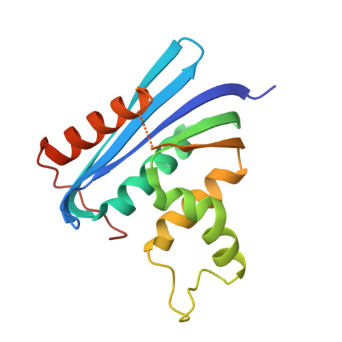
The Arg97 --> Gly and Asp136 --> His mutations stabilized So-RNase HI from the psychrotrophic bacterium Shewanella oneidensis MR-1 by 5.4 and 9.7 degrees C, respectively, in T(m), and 3.5 and 6.1 kJ x mol(-1), respectively, in DeltaG(H2O). These mutations also stabilized the So-RNase HI derivative (4x-RNase HI) with quadruple thermostabilizing mutations in an additive manner. As a result, the resultant sextuple mutant protein (6x-RNase HI) was more stable than the wild-type protein by 28.8 degrees C in T(m) and 27.0 kJ x mol(-1) in DeltaG(H2O). To analyse the effects of the mutations on the protein structure, the crystal structure of the 6x-RNase HI protein was determined at 2.5 A resolution. The main chain fold and interactions of the side-chains of the 6x-RNase HI protein were basically identical to those of the wild-type protein, except for the mutation sites. These results indicate that all six mutations independently affect the protein structure, and are consistent with the fact that the thermostabilizing effects of the mutations are roughly additive. The introduction of favourable interactions and the elimination of unfavourable interactions by the mutations contribute to the stabilization of the 6x-RNase HI protein. We propose that So-RNase HI is destabilized when compared with its mesophilic and thermophilic counterparts in a localized fashion by increasing the number of amino acid residues unfavourable for protein stability.
Organizational Affiliation:
Department of Material and Life Science, Graduate School of Engineering, Osaka University, Japan.