2ZOQ

Structural dissection of human mitogen-activated kinase ERK1


Experimental Data Snapshot

  • Method: X-RAY DIFFRACTION
  • Resolution: 2.39 Å
  • R-Value Free: 0.267 
  • R-Value Work: 0.249 

wwPDB Validation   3D Report Full Report


Ligand Structure Quality Assessment 


This is version 1.3 of the entry. See complete history


Literature

Crystal structure of human mono-phosphorylated ERK1 at Tyr204

Kinoshita, T.Yoshida, I.Nakae, S.Okita, K.Gouda, M.Matsubara, M.Yokota, K.Ishiguro, H.Tada, T.

(2008) Biochem Biophys Res Commun 377: 1123-1127

  • DOI: https://doi.org/10.1016/j.bbrc.2008.10.127
  • Primary Citation of Related Structures:  
    2ZOQ

  • PubMed Abstract: 

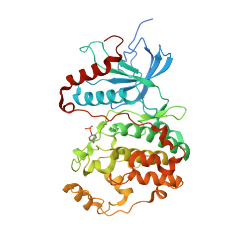
    Extracellular signal-regulated kinase (ERK) is a member of the MAP kinase family, and can regulate several cellular responses. The isoforms ERK1 and ERK2 have markedly similar amino acid sequences, but exhibit distinctive physiological functions. As well as ERK2, ERK1 was auto- and mono-phosphorylated at Tyr204 in the activation loop during Escherichia coli production, resulting in basal level activity, approximately 500-fold less compared with fully-active ERK1 dual-phosphorylated at Thr202 and Tyr204. Crystal structure demonstrated that the mono-phosphorylated ERK1 kinase possessed a novel conformation distinguishable from the un-phosphorylated (inactive) and the dual-phosphorylated (full-active) forms. The characteristic structural features in both the C-helix and the activation loop likely contribute to the basal activity of the mono-phosphorylated ERK1. The structural dissection of ERK1 compared to ERK2 suggests that the structural differences in the D-motif binding site and in the backside binding site are putative targets for development of selective ERK1/ERK2 inhibitors.


  • Organizational Affiliation

    Graduate School of Science, Osaka Prefecture University, 1-1 Gakuen-cho, Naka-ku, Sakai, Osaka 599-8531, Japan. kinotk@b.s.osakafu-u.ac.jp


Macromolecules
Find similar proteins by:  (by identity cutoff)  |  3D Structure
Entity ID: 1
MoleculeChains Sequence LengthOrganismDetailsImage
Mitogen-activated protein kinase 3
A, B
382Homo sapiensMutation(s): 0 
Gene Names: MAPK3ERK1PRKM3
EC: 2.7.11.24
UniProt & NIH Common Fund Data Resources
Find proteins for P27361 (Homo sapiens)
Explore P27361 
Go to UniProtKB:  P27361
PHAROS:  P27361
GTEx:  ENSG00000102882 
Entity Groups  
Sequence Clusters30% Identity50% Identity70% Identity90% Identity95% Identity100% Identity
UniProt GroupP27361
Sequence Annotations
Expand
  • Reference Sequence
Small Molecules
Modified Residues  1 Unique
IDChains TypeFormula2D DiagramParent
PTR
Query on PTR
A, B
L-PEPTIDE LINKINGC9 H12 N O6 PTYR
Binding Affinity Annotations 
IDSourceBinding Affinity
5ID BindingDB:  2ZOQ Ki: 530 (nM) from 1 assay(s)
IC50: 1200 (nM) from 1 assay(s)
Binding MOAD:  2ZOQ IC50: 900 (nM) from 1 assay(s)
PDBBind:  2ZOQ IC50: 900 (nM) from 1 assay(s)
Experimental Data & Validation

Experimental Data

  • Method: X-RAY DIFFRACTION
  • Resolution: 2.39 Å
  • R-Value Free: 0.267 
  • R-Value Work: 0.249 
  • Space Group: P 1 21 1
Unit Cell:
Length ( Å )Angle ( ˚ )
a = 62.382α = 90
b = 91.54β = 91.5
c = 64.93γ = 90
Software Package:
Software NamePurpose
ADSCdata collection
MOLREPphasing
CNXrefinement
HKL-2000data reduction
HKL-2000data scaling

Structure Validation

View Full Validation Report


Ligand Structure Quality Assessment 


Entry History 

Deposition Data

Revision History  (Full details and data files)

  • Version 1.0: 2009-04-07
    Type: Initial release
  • Version 1.1: 2011-07-13
    Changes: Version format compliance
  • Version 1.2: 2023-11-01
    Changes: Data collection, Database references, Derived calculations, Refinement description
  • Version 1.3: 2023-11-15
    Changes: Data collection