2ZLF

The Structural Basis for Peptidomimetic Inhibition of Eukaryotic Ribonucleotide Reductase


Experimental Data Snapshot

  • Method: X-RAY DIFFRACTION
  • Resolution: 2.59 Å
  • R-Value Free: 0.251 
  • R-Value Work: 0.192 
  • R-Value Observed: 0.196 

wwPDB Validation   3D Report Full Report


This is version 1.2 of the entry. See complete history


Literature

The Structural Basis for Peptidomimetic Inhibition of Eukaryotic Ribonucleotide Reductase: A Conformationally Flexible Pharmacophore

Xu, H.Fairman, J.W.Wijerathna, S.R.Kreischer, N.R.LaMacchia, J.Helmbrecht, E.Cooperman, B.S.Dealwis, C.

(2008) J Med Chem 51: 4653-4659

  • DOI: https://doi.org/10.1021/jm800350u
  • Primary Citation of Related Structures:  
    2ZLF, 2ZLG

  • PubMed Abstract: 

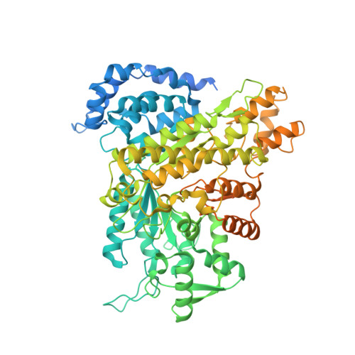
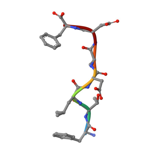
    Eukaryotic ribonucleotide reductase (RR) catalyzes nucleoside diphosphate conversion to deoxynucleoside diphosphate. Crucial for rapidly dividing cells, RR is a target for cancer therapy. RR activity requires formation of a complex between subunits R1 and R2 in which the R2 C-terminal peptide binds to R1. Here we report crystal structures of heterocomplexes containing mammalian R2 C-terminal heptapeptide, P7 (Ac-1FTLDADF7) and its peptidomimetic P6 (1Fmoc(Me)PhgLDChaDF7) bound to Saccharomyces cerevisiae R1 (ScR1). P7 and P6, both of which inhibit ScRR, each bind at two contiguous sites containing residues that are highly conserved among eukaryotes. Such binding is quite distinct from that reported for prokaryotes. The Fmoc group in P6 peptide makes several hydrophobic interactions that contribute to its enhanced potency in binding to ScR1. Combining all of our results, we observe three distinct conformations for peptide binding to ScR1. These structures provide pharmacophores for designing highly potent nonpeptide class I RR inhibitors.


  • Organizational Affiliation

    Department of Pharmacology, Case School of Medicine, Case Western Reserve University, 10900 Euclid Avenue, Cleveland, Ohio 44106-4965, USA.


Macromolecules
Find similar proteins by:  (by identity cutoff)  |  3D Structure
Entity ID: 1
MoleculeChains Sequence LengthOrganismDetailsImage
Ribonucleoside-diphosphate reductase large chain 1888Saccharomyces cerevisiaeMutation(s): 0 
EC: 1.17.4.1
UniProt
Find proteins for P21524 (Saccharomyces cerevisiae (strain ATCC 204508 / S288c))
Explore P21524 
Go to UniProtKB:  P21524
Entity Groups  
Sequence Clusters30% Identity50% Identity70% Identity90% Identity95% Identity100% Identity
UniProt GroupP21524
Sequence Annotations
Expand
  • Reference Sequence

Find similar proteins by:  Sequence   |   3D Structure  

Entity ID: 2
MoleculeChains Sequence LengthOrganismDetailsImage
FTLDADF7N/AMutation(s): 0 
Sequence Annotations
Expand
  • Reference Sequence
Experimental Data & Validation

Experimental Data

  • Method: X-RAY DIFFRACTION
  • Resolution: 2.59 Å
  • R-Value Free: 0.251 
  • R-Value Work: 0.192 
  • R-Value Observed: 0.196 
  • Space Group: P 21 21 2
Unit Cell:
Length ( Å )Angle ( ˚ )
a = 107.948α = 90
b = 116.999β = 90
c = 63.873γ = 90
Software Package:
Software NamePurpose
REFMACrefinement
HKL-2000data collection
HKL-2000data reduction
HKL-2000data scaling
CCP4phasing

Structure Validation

View Full Validation Report


Entry History 

Deposition Data

Revision History  (Full details and data files)

  • Version 1.0: 2008-08-19
    Type: Initial release
  • Version 1.1: 2011-07-13
    Changes: Version format compliance
  • Version 1.2: 2023-11-01
    Changes: Data collection, Database references, Refinement description