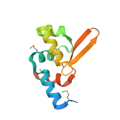
Crystal structure of the transcriptional repressor PagR of Bacillus anthracis
Zhao, H., Volkov, A., Veldore, V.H., Hoch, J.A., Varughese, K.I.(2010) Microbiology (N Y) 156: 385-391
- PubMed: 19926656
- DOI: https://doi.org/10.1099/mic.0.033548-0
- Primary Citation of Related Structures:
2ZKZ - PubMed Abstract:
PagR is a transcriptional repressor in Bacillus anthracis that controls the chromosomal S-layer genes eag and sap, and downregulates the protective antigen pagA gene by direct binding to their promoter regions. The PagR protein sequence is similar to those of members of the ArsR repressor family involved in the repression of arsenate-resistance genes in numerous bacteria. The crystal structure of PagR was solved using multi-wavelength anomalous diffraction (MAD) techniques and was refined with 1.8 A resolution diffraction data. The PagR molecules form dimers, as observed in all SmtB/ArsR repressor family proteins. In the crystal lattice four PagR dimers pack together to form an inactive octamer. Model-building studies suggest that the dimer binds to a DNA duplex with a bend of around 4 degrees.
Organizational Affiliation:
Department of Molecular and Experimental Medicine, The Scripps Research Institute, La Jolla, CA 92037, USA.