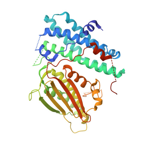
Pyruvate Dehydrogenase Kinase-4 Structures Reveal a Metastable Open Conformation Fostering Robust Core-free Basal Activity
Wynn, R.M., Kato, M., Chuang, J.L., Tso, S.-C., Li, J., Chuang, D.T.(2008) J Biol Chem 283: 25305-25315
- PubMed: 18658136
- DOI: https://doi.org/10.1074/jbc.M802249200
- Primary Citation of Related Structures:
2ZKJ, 3D2R - PubMed Abstract:
Human pyruvate dehydrogenase complex (PDC) is down-regulated by pyruvate dehydrogenase kinase (PDK) isoforms 1-4. PDK4 is overexpressed in skeletal muscle in type 2 diabetes, resulting in impaired glucose utilization. Here we show that human PDK4 has robust core-free basal activity, which is considerably higher than activity levels of other PDK isoforms stimulated by the PDC core. PDK4 binds the L3 lipoyl domain, but its activity is not significantly stimulated by any individual lipoyl domains or the core of PDC. The 2.0-A crystal structures of the PDK4 dimer with bound ADP reveal an open conformation with a wider active-site cleft, compared with that in the closed conformation epitomized by the PDK2-ADP structure. The open conformation in PDK4 shows partially ordered C-terminal cross-tails, in which the conserved DW (Asp(394)-Trp(395)) motif from one subunit anchors to the N-terminal domain of the other subunit. The open conformation fosters a reduced binding affinity for ADP, facilitating the efficient removal of product inhibition by this nucleotide. Alteration or deletion of the DW-motif disrupts the C-terminal cross-tail anchor, resulting in the closed conformation and the nearly complete inactivation of PDK4. Fluorescence quenching and enzyme activity data suggest that compounds AZD7545 and dichloroacetate lock PDK4 in the open and the closed conformational states, respectively. We propose that PDK4 with bound ADP exists in equilibrium between the open and the closed conformations. The favored metastable open conformation is responsible for the robust basal activity of PDK4 in the absence of the PDC core.
Organizational Affiliation:
Department of Biochemistry, Dallas, Texas 75390-9038; Department of Internal Medicine, University of Texas Southwestern Medical Center, Dallas, Texas 75390-9038.