2ZJ7

Crystal structure of D157A mutant of Pseudomonas sp. MIS38 lipase


Experimental Data Snapshot

  • Method: X-RAY DIFFRACTION
  • Resolution: 2.21 Å
  • R-Value Free: 0.234 
  • R-Value Work: 0.164 
  • R-Value Observed: 0.168 

wwPDB Validation   3D Report Full Report


This is version 1.3 of the entry. See complete history


Literature

Importance of the Ca2+-binding sites in the N-catalytic domain of a family I.3 lipase for activity and stability

Kuwahara, K.Angkawidjaja, C.Matsumura, H.Koga, Y.Takano, K.Kanaya, S.

(2008) Protein Eng Des Sel 21: 737-744

  • DOI: https://doi.org/10.1093/protein/gzn057
  • Primary Citation of Related Structures:  
    2ZJ6, 2ZJ7

  • PubMed Abstract: 

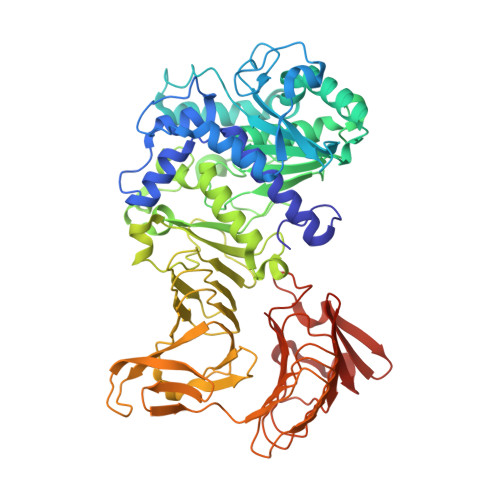
    A family I.3 lipase from Pseudomonas sp. MIS38 (PML) contains three Ca(2+)-binding sites (Ca1-Ca3) in the N-catalytic domain. Of them, the Ca1 site is formed only in an open conformation. To analyze the role of these Ca(2+)-binding sites, three mutant proteins D157A-PML, D275A-PML and D337A-PML, which are designed to remove the Ca1, Ca2 and Ca3 sites, respectively, were constructed. Of them, the crystal structures of D157A-PML and D337A-PML in a closed conformation were determined. Both structures are nearly identical to that of the wild-type protein, except that the Ca3 site is missing in the D337A-PML structure. D157A-PML was as stable as the wild-type protein. Nevertheless, it exhibited little lipase and very weak esterase activities. D275A-PML was less stable than the wild-type protein by approximately 5 degrees C in T(1/2). It exhibited weak but significant lipase and esterase activities when compared with the wild-type protein. D337A-PML was also less stable than the wild-type protein by approximately 5 degrees C in T(1/2) but was fully active. These results suggest that the Ca1 site is required to make the active site fully open by anchoring lid 1. The Ca2 and Ca3 sites contribute to the stabilization of PML. The Ca2 site is also required to make PML fully active.


  • Organizational Affiliation

    Department of Material and Life Science, Graduate School of Engineering, Osaka University, 2-1 Yamadaoka, Suita, Osaka 565-0871, Japan.


Macromolecules
Find similar proteins by:  (by identity cutoff)  |  3D Structure
Entity ID: 1
MoleculeChains Sequence LengthOrganismDetailsImage
Lipase617Pseudomonas sp. MIS38Mutation(s): 1 
EC: 3.1.1.3
UniProt
Find proteins for Q9RBY1 (Pseudomonas sp. MIS38)
Explore Q9RBY1 
Go to UniProtKB:  Q9RBY1
Entity Groups  
Sequence Clusters30% Identity50% Identity70% Identity90% Identity95% Identity100% Identity
UniProt GroupQ9RBY1
Sequence Annotations
Expand
  • Reference Sequence
Small Molecules
Ligands 2 Unique
IDChains Name / Formula / InChI Key2D Diagram3D Interactions
ZN
Query on ZN

Download Ideal Coordinates CCD File 
L [auth A],
M [auth A]
ZINC ION
Zn
PTFCDOFLOPIGGS-UHFFFAOYSA-N
CA
Query on CA

Download Ideal Coordinates CCD File 
B [auth A]
C [auth A]
D [auth A]
E [auth A]
F [auth A]
B [auth A],
C [auth A],
D [auth A],
E [auth A],
F [auth A],
G [auth A],
H [auth A],
I [auth A],
J [auth A],
K [auth A]
CALCIUM ION
Ca
BHPQYMZQTOCNFJ-UHFFFAOYSA-N
Experimental Data & Validation

Experimental Data

  • Method: X-RAY DIFFRACTION
  • Resolution: 2.21 Å
  • R-Value Free: 0.234 
  • R-Value Work: 0.164 
  • R-Value Observed: 0.168 
  • Space Group: P 1 21 1
Unit Cell:
Length ( Å )Angle ( ˚ )
a = 48.634α = 90
b = 84.627β = 96.85
c = 87.521γ = 90
Software Package:
Software NamePurpose
REFMACrefinement
HKL-2000data collection
HKL-2000data reduction
HKL-2000data scaling
MOLREPphasing

Structure Validation

View Full Validation Report


Entry History 

Deposition Data

Revision History  (Full details and data files)

  • Version 1.0: 2008-12-02
    Type: Initial release
  • Version 1.1: 2011-07-13
    Changes: Source and taxonomy, Version format compliance
  • Version 1.2: 2021-11-10
    Changes: Database references, Derived calculations
  • Version 1.3: 2023-11-01
    Changes: Data collection, Refinement description