2ZG1

Crystal Structure of Two N-terminal Domains of Siglec-5 in Complex with 6'-Sialyllactose


Experimental Data Snapshot

  • Method: X-RAY DIFFRACTION
  • Resolution: 2.70 Å
  • R-Value Free: 0.275 
  • R-Value Work: 0.246 

wwPDB Validation   3D Report Full Report


Ligand Structure Quality Assessment 


This is version 1.3 of the entry. See complete history


Literature

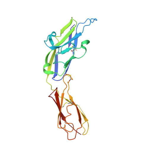
Structural implications of Siglec-5-mediated sialoglycan recognition

Zhuravleva, M.A.Trandem, K.Sun, P.D.

(2008) J Mol Biol 375: 437-447

  • DOI: https://doi.org/10.1016/j.jmb.2007.10.009
  • Primary Citation of Related Structures:  
    2ZG1, 2ZG2, 2ZG3

  • PubMed Abstract: 

    Sialic acid (Sia) Ig-like binding lectins are important mediators of recognition and signaling events among myeloid cells. To investigate the molecular mechanism underlying sialic acid Ig-like lectin (Siglec) functions, we determined the crystal structure of the two N-terminal extracellular domains of human myeloid cell inhibitory receptor Siglec-5 (CD170) and its complexes with two sialylated carbohydrates. The native structure revealed an unusual conformation of the CC' ligand specificity loop and a unique interdomain disulfide bond. The alpha(2,3)- and alpha(2,6)-sialyllactose complexed structures showed a conserved Sia recognition motif that involves both Arg124 and a portion of the G-strand in the V-set domain forming beta-sheet-like hydrogen bonds with the glycerol side chain of the Sia. Only few protein contacts to the subterminal sugars are observed and mediated by the highly variable GG' linker and CC' loop. These structural observations, in conjunction with surface plasmon resonance binding assays, provide mechanistic insights into linkage-dependent Siglec carbohydrate recognition and suggest that Siglec-5 and other CD33-related Siglec receptors are more promiscuous in sialoglycan recognition than previously understood.


  • Organizational Affiliation

    Structural Immunology Section, Laboratory of Immunogenetics, National Institute of Allergy and Infectious Diseases, National Institutes of Health, 12441 Parklawn Drive, Rockville, MD 20852, USA.


Macromolecules
Find similar proteins by:  (by identity cutoff)  |  3D Structure
Entity ID: 1
MoleculeChains Sequence LengthOrganismDetailsImage
Sialic acid-binding Ig-like lectin 5214Homo sapiensMutation(s): 0 
Gene Names: SIGLEC5
UniProt & NIH Common Fund Data Resources
Find proteins for O15389 (Homo sapiens)
Explore O15389 
Go to UniProtKB:  O15389
PHAROS:  O15389
Entity Groups  
Sequence Clusters30% Identity50% Identity70% Identity90% Identity95% Identity100% Identity
UniProt GroupO15389
Sequence Annotations
Expand
  • Reference Sequence
Small Molecules
Ligands 1 Unique
IDChains Name / Formula / InChI Key2D Diagram3D Interactions
SIA
Query on SIA

Download Ideal Coordinates CCD File 
B [auth A]N-acetyl-alpha-neuraminic acid
C11 H19 N O9
SQVRNKJHWKZAKO-YRMXFSIDSA-N
Binding Affinity Annotations 
IDSourceBinding Affinity
SIA PDBBind:  2ZG1 Kd: 8.00e+6 (nM) from 1 assay(s)
Experimental Data & Validation

Experimental Data

  • Method: X-RAY DIFFRACTION
  • Resolution: 2.70 Å
  • R-Value Free: 0.275 
  • R-Value Work: 0.246 
  • Space Group: H 3 2
Unit Cell:
Length ( Å )Angle ( ˚ )
a = 94.044α = 90
b = 94.044β = 90
c = 209.888γ = 120
Software Package:
Software NamePurpose
HKL-2000data collection
PHASERphasing
CNSrefinement
HKL-2000data reduction
SCALEPACKdata scaling

Structure Validation

View Full Validation Report


Ligand Structure Quality Assessment 


Entry History 

Deposition Data

Revision History  (Full details and data files)

  • Version 1.0: 2008-02-05
    Type: Initial release
  • Version 1.1: 2011-07-13
    Changes: Version format compliance
  • Version 1.2: 2020-07-29
    Type: Remediation
    Reason: Carbohydrate remediation
    Changes: Advisory, Data collection, Derived calculations, Structure summary
  • Version 1.3: 2023-11-01
    Changes: Advisory, Data collection, Database references, Refinement description, Structure summary