2ZFU

Structure of the methyltransferase-like domain of nucleomethylin


Experimental Data Snapshot

  • Method: X-RAY DIFFRACTION
  • Resolution: 2.00 Å
  • R-Value Free: 0.231 
  • R-Value Work: 0.192 
  • R-Value Observed: 0.194 

wwPDB Validation   3D Report Full Report


Ligand Structure Quality Assessment 


This is version 1.2 of the entry. See complete history


Literature

Epigenetic control of rDNA loci in response to intracellular energy status

Murayama, A.Ohmori, K.Fujimura, A.Minami, H.Yasuzawa-Tanaka, K.Kuroda, T.Oie, S.Daitoku, H.Okuwaki, M.Nagata, K.Fukamizu, A.Kimura, K.Shimizu, T.Yanagisawa, J.

(2008) Cell 133: 627-639

  • DOI: https://doi.org/10.1016/j.cell.2008.03.030
  • Primary Citation of Related Structures:  
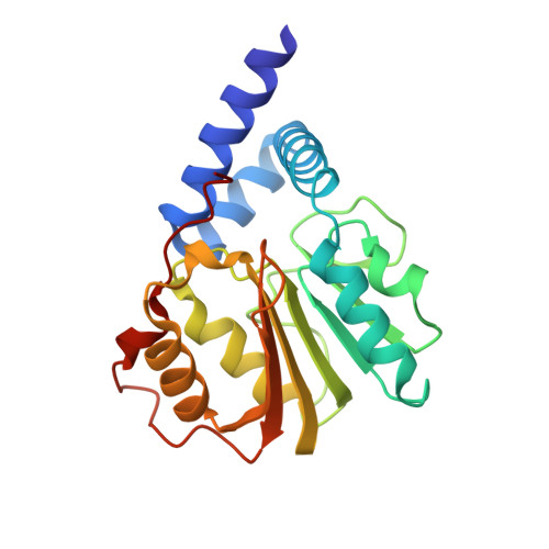
    2ZFU

  • PubMed Abstract: 

    Intracellular energy balance is important for cell survival. In eukaryotic cells, the most energy-consuming process is ribosome biosynthesis, which adapts to changes in intracellular energy status. However, the mechanism that links energy status and ribosome biosynthesis is largely unknown. Here, we describe eNoSC, a protein complex that senses energy status and controls rRNA transcription. eNoSC contains Nucleomethylin, which binds histone H3 dimethylated Lys9 in the rDNA locus, in a complex with SIRT1 and SUV39H1. Both SIRT1 and SUV39H1 are required for energy-dependent transcriptional repression, suggesting that a change in the NAD(+)/NADH ratio induced by reduction of energy status could activate SIRT1, leading to deacetylation of histone H3 and dimethylation at Lys9 by SUV39H1, thus establishing silent chromatin in the rDNA locus. Furthermore, eNoSC promotes restoration of energy balance by limiting rRNA transcription, thus protecting cells from energy deprivation-dependent apoptosis. These findings provide key insight into the mechanisms of energy homeostasis in cells.


  • Organizational Affiliation

    Graduate School of Life and Environmental Sciences, University of Tsukuba, 1-1-1 Tennodai, Tsukuba 305-8572, Japan.


Macromolecules
Find similar proteins by:  (by identity cutoff)  |  3D Structure
Entity ID: 1
MoleculeChains Sequence LengthOrganismDetailsImage
Cerebral protein 1
A, B
215Homo sapiensMutation(s): 0 
Gene Names: KIAA0409
UniProt & NIH Common Fund Data Resources
Find proteins for O43159 (Homo sapiens)
Explore O43159 
Go to UniProtKB:  O43159
PHAROS:  O43159
GTEx:  ENSG00000132275 
Entity Groups  
Sequence Clusters30% Identity50% Identity70% Identity90% Identity95% Identity100% Identity
UniProt GroupO43159
Sequence Annotations
Expand
  • Reference Sequence
Small Molecules
Experimental Data & Validation

Experimental Data

  • Method: X-RAY DIFFRACTION
  • Resolution: 2.00 Å
  • R-Value Free: 0.231 
  • R-Value Work: 0.192 
  • R-Value Observed: 0.194 
  • Space Group: P 61
Unit Cell:
Length ( Å )Angle ( ˚ )
a = 43.81α = 90
b = 43.81β = 90
c = 403.91γ = 120
Software Package:
Software NamePurpose
REFMACrefinement
ADSCdata collection
HKL-2000data reduction
HKL-2000data scaling
SOLVEphasing

Structure Validation

View Full Validation Report


Ligand Structure Quality Assessment 


Entry History 

Deposition Data

Revision History  (Full details and data files)

  • Version 1.0: 2008-12-02
    Type: Initial release
  • Version 1.1: 2011-07-13
    Changes: Advisory, Version format compliance
  • Version 1.2: 2024-03-13
    Changes: Data collection, Database references, Derived calculations