2ZBK

Crystal structure of an intact type II DNA topoisomerase: insights into DNA transfer mechanisms


Experimental Data Snapshot

  • Method: X-RAY DIFFRACTION
  • Resolution: 3.56 Å
  • R-Value Free: 0.333 
  • R-Value Work: 0.312 
  • R-Value Observed: 0.313 

wwPDB Validation   3D Report Full Report


Ligand Structure Quality Assessment 


This is version 1.2 of the entry. See complete history


Literature

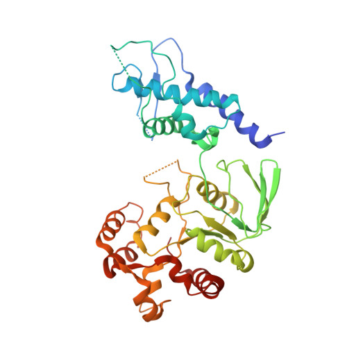
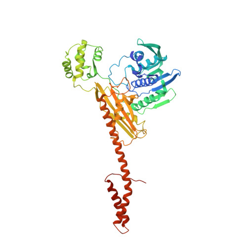
Crystal Structure of an Intact Type II DNA Topoisomerase: Insights into DNA Transfer Mechanisms

Graille, M.Durand, D.Lecointe, F.Gadelle, D.Quevillon-Cheruel, S.Vachette, P.Forterre, P.van Tilbeurgh, H.

(2008) Structure 16: 360-370

  • DOI: https://doi.org/10.1016/j.str.2007.12.020
  • Primary Citation of Related Structures:  
    2ZBK

  • PubMed Abstract: 

    DNA topoisomerases resolve DNA topological problems created during transcription, replication, and recombination. These ubiquitous enzymes are essential for cell viability and are highly potent targets for the development of antibacterial and antitumoral drugs. Type II enzymes catalyze the transfer of a DNA duplex through another one in an ATP-dependent mechanism. Because of its small size and sensitivity to antitumoral drugs, the archaeal DNA topoisomerase VI, a type II enzyme, is an excellent model for gaining further understanding of the organization and mechanism of these enzymes. We present the crystal structure of intact DNA topoisomerase VI bound to radicicol, an inhibitor of human topo II, and compare it to the conformation of the apo-protein as determined by small-angle X-ray scattering in solution. This structure, combined with a wealth of experimental data gathered on these enzymes, allows us to propose a structural model for the two-gate DNA transfer mechanism.


  • Organizational Affiliation

    Institut de Biochimie et de Biophysique Moléculaire et Cellulaire, UMR8619 CNRS, Université Paris-Sud, IFR115, F-91405 Orsay, France.


Macromolecules
Find similar proteins by:  (by identity cutoff)  |  3D Structure
Entity ID: 1
MoleculeChains Sequence LengthOrganismDetailsImage
Type II DNA topoisomerase VI subunit A
A, C, E, G
389Saccharolobus shibataeMutation(s): 0 
Gene Names: top6A
EC: 5.99.1.3
UniProt
Find proteins for O05208 (Saccharolobus shibatae (strain ATCC 51178 / DSM 5389 / JCM 8931 / NBRC 15437 / B12))
Explore O05208 
Go to UniProtKB:  O05208
Entity Groups  
Sequence Clusters30% Identity50% Identity70% Identity90% Identity95% Identity100% Identity
UniProt GroupO05208
Sequence Annotations
Expand
  • Reference Sequence
Find similar proteins by:  (by identity cutoff)  |  3D Structure
Entity ID: 2
MoleculeChains Sequence LengthOrganismDetailsImage
Type 2 DNA topoisomerase 6 subunit B
B, D, F, H
530Saccharolobus shibataeMutation(s): 0 
Gene Names: top6B
EC: 5.99.1.3
UniProt
Find proteins for O05207 (Saccharolobus shibatae (strain ATCC 51178 / DSM 5389 / JCM 8931 / NBRC 15437 / B12))
Explore O05207 
Go to UniProtKB:  O05207
Entity Groups  
Sequence Clusters30% Identity50% Identity70% Identity90% Identity95% Identity100% Identity
UniProt GroupO05207
Sequence Annotations
Expand
  • Reference Sequence
Experimental Data & Validation

Experimental Data

  • Method: X-RAY DIFFRACTION
  • Resolution: 3.56 Å
  • R-Value Free: 0.333 
  • R-Value Work: 0.312 
  • R-Value Observed: 0.313 
  • Space Group: P 21 21 21
Unit Cell:
Length ( Å )Angle ( ˚ )
a = 114.96α = 90
b = 200.53β = 90
c = 329.07γ = 90
Software Package:
Software NamePurpose
XSCALEdata scaling
SHELXphasing
REFMACrefinement
PDB_EXTRACTdata extraction
ADSCdata collection
SHELXDphasing

Structure Validation

View Full Validation Report


Ligand Structure Quality Assessment 


Entry History 

Deposition Data

Revision History  (Full details and data files)

  • Version 1.0: 2008-02-12
    Type: Initial release
  • Version 1.1: 2011-07-13
    Changes: Version format compliance
  • Version 1.2: 2023-11-01
    Changes: Data collection, Database references, Derived calculations, Refinement description