2Z6M

Crystal structure of Human Ferritin H8 as biotemplate for noble metal nanoparticle synthesis


Experimental Data Snapshot

  • Method: X-RAY DIFFRACTION
  • Resolution: 2.72 Å
  • R-Value Free: 0.252 
  • R-Value Work: 0.215 

wwPDB Validation   3D Report Full Report


This is version 1.3 of the entry. See complete history


Literature

Directing noble metal ion chemistry within a designed ferritin protein

Butts, C.A.Swift, J.Kang, S.G.Di Costanzo, L.Christianson, D.W.Saven, J.G.Dmochowski, I.J.

(2008) Biochemistry 47: 12729-12739

  • DOI: https://doi.org/10.1021/bi8016735
  • Primary Citation of Related Structures:  
    2Z6M, 3ERZ, 3ES3

  • PubMed Abstract: 

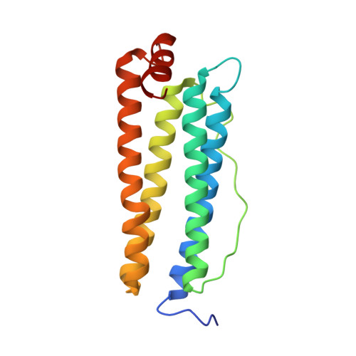
    Human H ferritin (HuHF) assembles from 24 four-helix bundles to form an approximately 500 kDa protein with an 8 nm internal cavity. HuHF provides a useful model for studying the transport of metal ions in solution to buried reaction sites in proteins. In this study, HuHF was redesigned to facilitate noble metal ion (Au(3+), Ag(+)) binding, reduction, and nanoparticle formation within the cavity. Computationally determined amino acid substitutions were targeted at four external and four internal surface sites. A variant with a total of 96 cysteines and histidines removed from the exterior surface and 96 non-native cysteines added to the interior surface retained wild-type stability and structure, as confirmed by X-ray crystallography, and promoted the formation of silver or gold nanoparticles within the protein cavity. Crystallographic studies with HuHF variants provide insight into how ferritins control access of metal ions to interior residues that perform chemistry.


  • Organizational Affiliation

    Department of Chemistry, University of Pennsylvania, 231 South 34th Street, Philadelphia, Pennsylvania 19104-6323, USA.


Macromolecules
Find similar proteins by:  (by identity cutoff)  |  3D Structure
Entity ID: 1
MoleculeChains Sequence LengthOrganismDetailsImage
Ferritin heavy chain
A, B, C, D, E
A, B, C, D, E, F, G, H, I, J, K, L
176Homo sapiensMutation(s): 8 
EC: 1.16.3.1
UniProt & NIH Common Fund Data Resources
Find proteins for P02794 (Homo sapiens)
Explore P02794 
Go to UniProtKB:  P02794
PHAROS:  P02794
GTEx:  ENSG00000167996 
Entity Groups  
Sequence Clusters30% Identity50% Identity70% Identity90% Identity95% Identity100% Identity
UniProt GroupP02794
Sequence Annotations
Expand
  • Reference Sequence
Small Molecules
Ligands 3 Unique
IDChains Name / Formula / InChI Key2D Diagram3D Interactions
MPD
Query on MPD

Download Ideal Coordinates CCD File 
AA [auth I]
BA [auth L]
Q [auth B]
R [auth C]
S [auth E]
AA [auth I],
BA [auth L],
Q [auth B],
R [auth C],
S [auth E],
T [auth F],
Y [auth H]
(4S)-2-METHYL-2,4-PENTANEDIOL
C6 H14 O2
SVTBMSDMJJWYQN-YFKPBYRVSA-N
ZN
Query on ZN

Download Ideal Coordinates CCD File 
M [auth A],
O [auth B],
U [auth G],
W [auth H],
Z [auth I]
ZINC ION
Zn
PTFCDOFLOPIGGS-UHFFFAOYSA-N
CA
Query on CA

Download Ideal Coordinates CCD File 
N [auth A],
P [auth B],
V [auth G],
X [auth H]
CALCIUM ION
Ca
BHPQYMZQTOCNFJ-UHFFFAOYSA-N
Experimental Data & Validation

Experimental Data

  • Method: X-RAY DIFFRACTION
  • Resolution: 2.72 Å
  • R-Value Free: 0.252 
  • R-Value Work: 0.215 
  • Space Group: P 4 21 2
Unit Cell:
Length ( Å )Angle ( ˚ )
a = 171.294α = 90
b = 171.294β = 90
c = 190.455γ = 90
Software Package:
Software NamePurpose
ADSCdata collection
PHASERphasing
CNSrefinement
HKL-2000data reduction
SCALEPACKdata scaling

Structure Validation

View Full Validation Report


Entry History 

Deposition Data

Revision History  (Full details and data files)

  • Version 1.0: 2008-07-22
    Type: Initial release
  • Version 1.1: 2011-07-13
    Changes: Version format compliance
  • Version 1.2: 2021-11-10
    Changes: Database references, Derived calculations
  • Version 1.3: 2023-11-01
    Changes: Data collection, Refinement description