2Z5S

Molecular basis for the inhibition of p53 by Mdmx


Experimental Data Snapshot

  • Method: X-RAY DIFFRACTION
  • Resolution: 2.30 Å
  • R-Value Free: 0.310 
  • R-Value Work: 0.247 
  • R-Value Observed: 0.251 

wwPDB Validation   3D Report Full Report


This is version 1.3 of the entry. See complete history


Literature

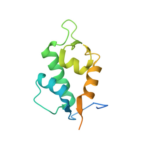
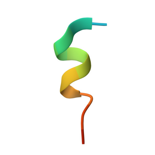
Molecular basis for the inhibition of p53 by Mdmx.

Popowicz, G.M.Czarna, A.Rothweiler, U.Szwagierczak, A.Krajewski, M.Weber, L.Holak, T.A.

(2007) Cell Cycle 6: 2386-2392

  • DOI: https://doi.org/10.4161/cc.6.19.4740
  • Primary Citation of Related Structures:  
    2Z5S, 2Z5T

  • PubMed Abstract: 

    The oncoprotein Mdm2, and the recently intensely studied, homologues protein Mdmx, are principal negative regulators of the p53 tumor suppressor. The mechanisms by which they regulate the stability and activity of p53 are not fully established. We have determined the crystal structure of the N-terminal domain of Mdmx bound to a 15-residue p53 peptide. The structure reveals that although the principle features of the Mdm2-p53 interaction are preserved in the Mdmx-p53 complex, the Mdmx hydrophobic cleft on which the p53 peptide binds is significantly altered: a part of the cleft is blocked by sidechains of Met and Tyr of the p53-binding pocket of Mdmx. Thus specific inhibitors of Mdm2-p53 would not be optimal for binding to Mdmx. Our binding assays show indeed that nutlins, the newly discovered, potent antagonists of the Mdm2-p53 interaction, are not capable to efficiently disrupt the Mdmx-p53 interaction. To achieve full activation of p53 in tumor cells, compounds that are specific for Mdmx are necessary to complement the Mdm2 specific binders.


  • Organizational Affiliation

    Max Planck Institute for Biochemistry, Martinsried, Germany.


Macromolecules
Find similar proteins by:  (by identity cutoff)  |  3D Structure
Entity ID: 1
MoleculeChains Sequence LengthOrganismDetailsImage
Mdm4 proteinA [auth M],
C [auth N],
E [auth O]
140Danio rerioMutation(s): 0 
Gene Names: mdm4
UniProt
Find proteins for Q7ZUW7 (Danio rerio)
Explore Q7ZUW7 
Go to UniProtKB:  Q7ZUW7
Entity Groups  
Sequence Clusters30% Identity50% Identity70% Identity90% Identity95% Identity100% Identity
UniProt GroupQ7ZUW7
Sequence Annotations
Expand
  • Reference Sequence

Find similar proteins by:  Sequence   |   3D Structure  

Entity ID: 2
MoleculeChains Sequence LengthOrganismDetailsImage
Cellular tumor antigen p53B [auth P],
D [auth Q],
F [auth R]
15N/AMutation(s): 0 
UniProt & NIH Common Fund Data Resources
Find proteins for P04637 (Homo sapiens)
Explore P04637 
Go to UniProtKB:  P04637
PHAROS:  P04637
GTEx:  ENSG00000141510 
Entity Groups  
Sequence Clusters30% Identity50% Identity70% Identity90% Identity95% Identity100% Identity
UniProt GroupP04637
Sequence Annotations
Expand
  • Reference Sequence
Experimental Data & Validation

Experimental Data

  • Method: X-RAY DIFFRACTION
  • Resolution: 2.30 Å
  • R-Value Free: 0.310 
  • R-Value Work: 0.247 
  • R-Value Observed: 0.251 
  • Space Group: P 1 21 1
Unit Cell:
Length ( Å )Angle ( ˚ )
a = 80.52α = 90
b = 30.44β = 102.47
c = 100.85γ = 90
Software Package:
Software NamePurpose
REFMACrefinement
MAR345dtbdata collection
XDSdata reduction
XSCALEdata scaling
MOLREPphasing

Structure Validation

View Full Validation Report


Entry History 

Deposition Data

Revision History  (Full details and data files)

  • Version 1.0: 2007-11-06
    Type: Initial release
  • Version 1.1: 2011-07-13
    Changes: Version format compliance
  • Version 1.2: 2019-11-06
    Changes: Data collection, Database references
  • Version 1.3: 2023-11-01
    Changes: Data collection, Database references, Refinement description