


Crystal structure of the human Phosphodiesterase 9A catalytic domain
Handa, N., Shirouzu, M., Terada, T., Omori, K., Kotera, J., Yokoyama, S.To be published.
Experimental Data Snapshot
Entity ID: 1 | |||||
|---|---|---|---|---|---|
| Molecule | Chains | Sequence Length | Organism | Details | Image |
| High-affinity cGMP-specific 3',5'-cyclic phosphodiesterase 9A | 333 | Homo sapiens | Mutation(s): 0 EC: 3.1.4.35 | 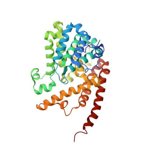 | |
UniProt & NIH Common Fund Data Resources | |||||
Find proteins for O76083 (Homo sapiens) Explore O76083 Go to UniProtKB: O76083 | |||||
PHAROS: O76083 GTEx: ENSG00000160191 | |||||
Entity Groups | |||||
| Sequence Clusters | 30% Identity50% Identity70% Identity90% Identity95% Identity100% Identity | ||||
| UniProt Group | O76083 | ||||
Sequence AnnotationsExpand | |||||
| |||||
| Ligands 3 Unique | |||||
|---|---|---|---|---|---|
| ID | Chains | Name / Formula / InChI Key | 2D Diagram | 3D Interactions | |
| IBM Query on IBM | E [auth A], H [auth B] | 3-ISOBUTYL-1-METHYLXANTHINE C10 H14 N4 O2 APIXJSLKIYYUKG-UHFFFAOYSA-N |  | ||
| ZN Query on ZN | C [auth A], F [auth B] | ZINC ION Zn PTFCDOFLOPIGGS-UHFFFAOYSA-N |  | ||
| MG Query on MG | D [auth A], G [auth B] | MAGNESIUM ION Mg JLVVSXFLKOJNIY-UHFFFAOYSA-N |  | ||
| Length ( Å ) | Angle ( ˚ ) |
|---|---|
| a = 104.27 | α = 90 |
| b = 104.27 | β = 90 |
| c = 269.61 | γ = 90 |
| Software Name | Purpose |
|---|---|
| CNS | refinement |
| HKL-2000 | data collection |
| HKL-2000 | data reduction |
| HKL-2000 | data scaling |
| MOLREP | phasing |
RCSB PDB (citation) is hosted by
RCSB PDB is a member of the

