2YXL

Crystal Structure of PH0851


Experimental Data Snapshot

  • Method: X-RAY DIFFRACTION
  • Resolution: 2.55 Å
  • R-Value Free: 0.246 
  • R-Value Work: 0.212 
  • R-Value Observed: 0.214 

wwPDB Validation   3D Report Full Report


Ligand Structure Quality Assessment 


This is version 1.2 of the entry. See complete history


Literature

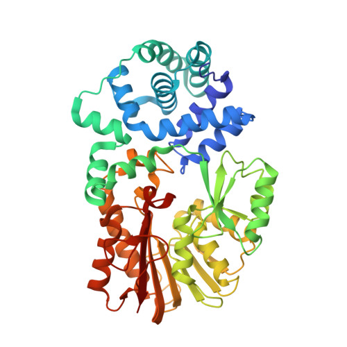
Structure of an archaeal homologue of the bacterial Fmu/RsmB/RrmB rRNA cytosine 5-methyltransferase

Hikida, Y.Kuratani, M.Bessho, Y.Sekine, S.Yokoyama, S.

(2010) Acta Crystallogr D Biol Crystallogr 66: 1301-1307

  • DOI: https://doi.org/10.1107/S0907444910037558
  • Primary Citation of Related Structures:  
    2YXL

  • PubMed Abstract: 

    One of the modified nucleosides that frequently occurs in rRNAs and tRNAs is 5-methylcytidine (m⁵C). Escherichia coli Fmu/RsmB/RrmB is an S-adenosyl-L-methionine (AdoMet)-dependent methyltransferase that forms m⁵C967 in 16S rRNA. Fmu/RsmB/RrmB homologues exist not only in bacteria but also in archaea and eukarya and constitute a large orthologous group in the RNA:m⁵C methyltransferase family. In the present study, the crystal structure of a homologue of E. coli Fmu/RsmB/RrmB from the archaeon Pyrococcus horikoshii (PH0851) complexed with an AdoMet analogue was determined at 2.55 Å resolution. The structure and sequence of the C-terminal catalytic domain are highly conserved compared with those of E. coli Fmu/RsmB/RrmB. In contrast, the sequence of the N-terminal domain is negligibly conserved between the bacterial and archaeal subfamilies. Nevertheless, the N-terminal domains of PH0851 and E. coli Fmu/RsmB/RrmB are both α-helical and adopt a similar topology. Next to the AdoMet-binding site, a positively charged cleft is formed between the N- and C-terminal domains. This cleft is conserved in the archaeal PH0851 homologues and seems to be suitable for binding the RNA substrate.


  • Organizational Affiliation

    Department of Biophysics and Biochemistry, Graduate School of Science, The University of Tokyo, 7-3-1 Hongo, Bunkyo-ku, Tokyo 113-0033, Japan.


Macromolecules
Find similar proteins by:  (by identity cutoff)  |  3D Structure
Entity ID: 1
MoleculeChains Sequence LengthOrganismDetailsImage
450aa long hypothetical fmu protein450Pyrococcus horikoshii OT3Mutation(s): 0 
Gene Names: PH0851
UniProt
Find proteins for O58581 (Pyrococcus horikoshii (strain ATCC 700860 / DSM 12428 / JCM 9974 / NBRC 100139 / OT-3))
Explore O58581 
Go to UniProtKB:  O58581
Entity Groups  
Sequence Clusters30% Identity50% Identity70% Identity90% Identity95% Identity100% Identity
UniProt GroupO58581
Sequence Annotations
Expand
  • Reference Sequence
Small Molecules
Ligands 1 Unique
IDChains Name / Formula / InChI Key2D Diagram3D Interactions
SFG
Query on SFG

Download Ideal Coordinates CCD File 
B [auth A]SINEFUNGIN
C15 H23 N7 O5
LMXOHSDXUQEUSF-YECHIGJVSA-N
Experimental Data & Validation

Experimental Data

  • Method: X-RAY DIFFRACTION
  • Resolution: 2.55 Å
  • R-Value Free: 0.246 
  • R-Value Work: 0.212 
  • R-Value Observed: 0.214 
  • Space Group: P 32 2 1
Unit Cell:
Length ( Å )Angle ( ˚ )
a = 116.621α = 90
b = 116.621β = 90
c = 93.684γ = 120
Software Package:
Software NamePurpose
REFMACrefinement
HKL-2000data collection
HKL-2000data reduction
HKL-2000data scaling
MOLREPphasing

Structure Validation

View Full Validation Report


Ligand Structure Quality Assessment 


Entry History 

Deposition Data

Revision History  (Full details and data files)

  • Version 1.0: 2008-04-29
    Type: Initial release
  • Version 1.1: 2011-07-13
    Changes: Advisory, Source and taxonomy, Version format compliance
  • Version 1.2: 2023-10-25
    Changes: Data collection, Database references, Derived calculations, Refinement description