


Crystal structure of peroxiredoxin-like protein from Sulfolobus tokodaii
Ebihara, A., Manzoku, M., Fujimoto, Y., Yokoyama, S., Kuramitsu, S.To be published.
Experimental Data Snapshot
wwPDB Validation 3D Report Full Report

A newer entry is available that reflects an alternative modeling of the original data: 4G2E
Entity ID: 1 | |||||
|---|---|---|---|---|---|
| Molecule | Chains | Sequence Length | Organism | Details | Image |
| Peroxiredoxin-like protein | 157 | Sulfurisphaera tokodaii | Mutation(s): 0 | 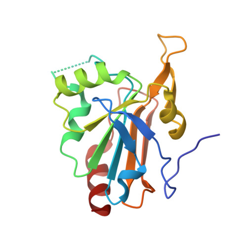 | |
UniProt | |||||
Find proteins for F9VNL8 (Sulfurisphaera tokodaii (strain DSM 16993 / JCM 10545 / NBRC 100140 / 7)) Explore F9VNL8 Go to UniProtKB: F9VNL8 | |||||
Entity Groups | |||||
| Sequence Clusters | 30% Identity50% Identity70% Identity90% Identity95% Identity100% Identity | ||||
| UniProt Group | F9VNL8 | ||||
Sequence AnnotationsExpand | |||||
| |||||
| Length ( Å ) | Angle ( ˚ ) |
|---|---|
| a = 69.34 | α = 90 |
| b = 78.67 | β = 90 |
| c = 61.97 | γ = 90 |
| Software Name | Purpose |
|---|---|
| CNS | refinement |
| HKL-2000 | data reduction |
| HKL-2000 | data scaling |
| MOLREP | phasing |
RCSB PDB (citation) is hosted by
RCSB PDB is a member of the

