2YMV

Structure of Reduced M Smegmatis 5246, a homologue of M.Tuberculosis Acg


Experimental Data Snapshot

  • Method: X-RAY DIFFRACTION
  • Resolution: 1.60 Å
  • R-Value Free: 0.147 
  • R-Value Work: 0.119 
  • R-Value Observed: 0.120 

wwPDB Validation   3D Report Full Report


Ligand Structure Quality Assessment 


This is version 1.1 of the entry. See complete history


Literature

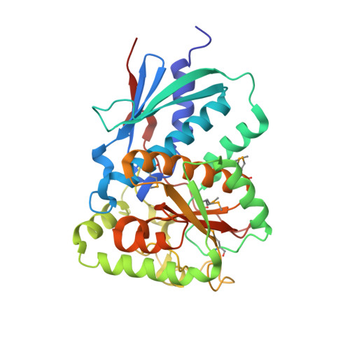
Crystal Structure of Reduced Msacg, a Putative Nitroreductase from Mycobacterium Smegmatis and a Close Homologue of Mycobacterium Tuberculosis Acg.

Chauviac, F.-X.Bommer, M.Yan, J.Parkin, G.Daviter, T.Lowden, P.Raven, E.L.Thalassinos, K.Keep, N.H.

(2012) J Biol Chem 287: 44372

  • DOI: https://doi.org/10.1074/jbc.M112.406264
  • Primary Citation of Related Structures:  
    2YMV

  • PubMed Abstract: 

    This paper presents the structure of MsAcg (MSMEG_5246), a Mycobacterium smegmatis homologue of Mycobacterium tuberculosis Acg (Rv2032) in its reduced form at 1.6 Å resolution using x-ray crystallography. Rv2032 is one of the most induced genes under the hypoxic model of tuberculosis dormancy. The Acg family turns out to be unusual flavin mononucleotide (FMN)-binding proteins that have probably arisen by gene duplication and fusion from a classical homodimeric nitroreductase such that the monomeric protein resembles a classical nitroreductase dimer but with one active site deleted and the other active site covered by a unique lid. The FMN cofactor is not reduced by either NADH or NADPH, but the chemically reduced enzyme is capable of reduction of nitro substrates, albeit at no kinetic advantage over free FMN. The reduced enzyme is rapidly oxidized by oxygen but without any evidence for a radical state commonly seen in oxygen-sensitive nitroreductases. The presence of the unique lid domain, the lack of reduction by NAD(P)H, and the slow rate of reaction of the chemically reduced protein raises a possible alternative function of Acg proteins in FMN storage or sequestration from other biochemical pathways as part of the bacteria's adaptation to a dormancy state.


  • Organizational Affiliation

    Crystallography, Institute for Structural and Molecular Biology, Department of Biological Sciences, Birkbeck, University of London, Malet Street, London, WC1E 7HX, United Kingdom.


Macromolecules
Find similar proteins by:  (by identity cutoff)  |  3D Structure
Entity ID: 1
MoleculeChains Sequence LengthOrganismDetailsImage
ACG NITROREDUCTASE330Mycolicibacterium smegmatisMutation(s): 0 
UniProt
Find proteins for A0R2V4 (Mycolicibacterium smegmatis (strain ATCC 700084 / mc(2)155))
Explore A0R2V4 
Go to UniProtKB:  A0R2V4
Entity Groups  
Sequence Clusters30% Identity50% Identity70% Identity90% Identity95% Identity100% Identity
UniProt GroupA0R2V4
Sequence Annotations
Expand
  • Reference Sequence
Small Molecules
Ligands 5 Unique
IDChains Name / Formula / InChI Key2D Diagram3D Interactions
FNR
Query on FNR

Download Ideal Coordinates CCD File 
B [auth A]1-DEOXY-1-(7,8-DIMETHYL-2,4-DIOXO-3,4-DIHYDRO-2H-BENZO[G]PTERIDIN-1-ID-10(5H)-YL)-5-O-PHOSPHONATO-D-RIBITOL
C17 H23 N4 O9 P
YTNIXZGTHTVJBW-SCRDCRAPSA-N
PGE
Query on PGE

Download Ideal Coordinates CCD File 
I [auth A],
J [auth A]
TRIETHYLENE GLYCOL
C6 H14 O4
ZIBGPFATKBEMQZ-UHFFFAOYSA-N
IMD
Query on IMD

Download Ideal Coordinates CCD File 
D [auth A]IMIDAZOLE
C3 H5 N2
RAXXELZNTBOGNW-UHFFFAOYSA-O
ACT
Query on ACT

Download Ideal Coordinates CCD File 
F [auth A],
H [auth A]
ACETATE ION
C2 H3 O2
QTBSBXVTEAMEQO-UHFFFAOYSA-M
FMT
Query on FMT

Download Ideal Coordinates CCD File 
C [auth A],
E [auth A],
G [auth A]
FORMIC ACID
C H2 O2
BDAGIHXWWSANSR-UHFFFAOYSA-N
Modified Residues  1 Unique
IDChains TypeFormula2D DiagramParent
MSE
Query on MSE
A
L-PEPTIDE LINKINGC5 H11 N O2 SeMET
Experimental Data & Validation

Experimental Data

  • Method: X-RAY DIFFRACTION
  • Resolution: 1.60 Å
  • R-Value Free: 0.147 
  • R-Value Work: 0.119 
  • R-Value Observed: 0.120 
  • Space Group: P 3 2 1
Unit Cell:
Length ( Å )Angle ( ˚ )
a = 167.799α = 90
b = 167.799β = 90
c = 43.98γ = 120
Software Package:
Software NamePurpose
PHENIXrefinement
XDSdata reduction
Aimlessdata scaling
PHASERphasing

Structure Validation

View Full Validation Report


Ligand Structure Quality Assessment 


Entry History 

Deposition Data

Revision History  (Full details and data files)

  • Version 1.0: 2012-11-21
    Type: Initial release
  • Version 1.1: 2013-01-16
    Changes: Database references