Crystal Structure of Sol I 2, a Major Allergen from Fire Ant Venom
Borer, A.S., Wassmann, P., Schmidt, M., Hoffman, D.R., Zhou, J.J., Wright, C., Schirmer, T., Markovic-Housley, Z.(2012) J Mol Biol 415: 635
- PubMed: 22100449
- DOI: https://doi.org/10.1016/j.jmb.2011.10.009
- Primary Citation of Related Structures:
2YGU - PubMed Abstract:
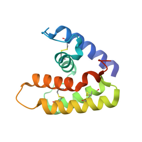
Sol i 2 is a potent allergen from the venom of red imported fire ant, which contains allergens Sol i 1, Sol i 2, Sol i 3, and Sol i 4 that are known to be powerful triggers of anaphylaxis. Sol i 2 causes IgE antibody production in about one-third of individuals stung by fire ants. Baculovirus recombinant dimeric Sol i 2 was crystallized as a native and selenomethionyl-derivatized protein, and its structure has been determined by single-wavelength anomalous dispersion at 2.6 Å resolution. The overall fold of each subunit consists of five helices that enclose a central hydrophobic cavity. The structure is stabilized by three intramolecular disulfide bridges and one intermolecular disulfide bridge. The nearest structural homologue is the sequence-unrelated odorant binding protein and pheromone binding protein LUSH of the fruit fly Drosophila, which may suggest a similar biological function. To test this hypothesis, we measured the reversible binding of various pheromones, plant odorants, and other ligands to Sol i 2 by the changes in N-phenyl-1-naphthylamine fluorescence emission upon binding of ligands that compete with N-phenyl-1-naphthylamine. The highest binding affinity was observed for hydrophobic ligands such as aphid alarm pheromone (E)-β-farnesene, analogs of ant alarm pheromones, and plant volatiles decane, undecane, and β-caryophyllene. Conceivably, Sol i 2 may play a role in capturing and/or transporting small hydrophobic ligands such as pheromones, odors, fatty acids, or short-living hydrophobic primers. Molecular surface analysis, in combination with sequence alignment, can explain the serological cross-reactivity observed between some ant species.
Organizational Affiliation:
Department of Structural Biology, Biozentrum, University of Basel, Basel, Switzerland.