The Overall Architecture and Receptor Binding of Pneumococcal Carbohydrate Antigen Hydrolyzing Enzymes.
Higgins, M.A., Ficko-Blean, E., Wright, C., Meloncelli, P.J., Lowary, T.L., Boraston, A.B.(2011) J Mol Biol 411: 1017
- PubMed: 21767550
- DOI: https://doi.org/10.1016/j.jmb.2011.06.035
- Primary Citation of Related Structures:
2YGL, 2YGM - PubMed Abstract:
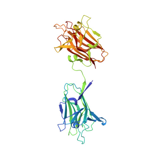
The TIGR4 and SP3-BS71 strains of Streptococcus pneumoniae each produce family 98 glycoside hydrolases, called Sp4GH98 and Sp3GH98, respectively, which have different modular architectures and substrate specificities. Sp4GH98 degrades the Lewis(Y) antigen and possesses three C-terminal family 47 carbohydrate-binding modules (CBMs) that bind to this substrate. Sp3GH98 degrades the blood group A/B antigens and has two N-terminal family 51 CBMs that are of unknown function. Here, we examine the complex carbohydrate-binding specificity of the family 51 CBMs from Sp3GH98 (referred to as CBM51-1 and CBM51-2), the structural basis of this interaction, and the overall solution conformations of both Sp3GH98 and Sp4GH98, which are shown to be fully secreted proteins. Through glycan microarray binding analysis and isothermal titration calorimetry, CBM51-1 is found to bind specifically to the blood group A/B antigens. However, due to a series of relatively small structural rearrangements that were revealed in structures determined by X-ray crystallography, CBM51-2 appears to be incapable of binding carbohydrates. Analysis of small-angle X-ray scattering data in combination with the available high-resolution X-ray crystal structures of the Sp3GH98 and Sp4GH98 catalytic modules and their CBMs yielded models of the biological solution structures of the full-length enzymes. These studies reveal the complex architectures of the two enzymes and suggest that carbohydrate recognition by the CBMs and the activity of the catalytic modules are not directly coupled.
Organizational Affiliation:
Biochemistry and Microbiology, University of Victoria, PO Box 3055 STN CSC, Victoria, BC, Canada V8W 3P6.