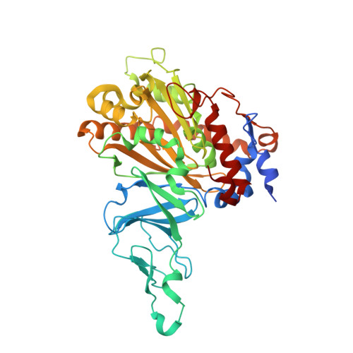
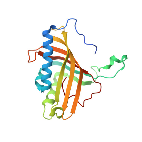
2YFL

Crystal Structure of Biphenyl dioxygenase variant RR41 with 2-chloro dibenzofuran


Experimental Data Snapshot

  • Method: X-RAY DIFFRACTION
  • Resolution: 2.60 Å
  • R-Value Free: 0.280 
  • R-Value Work: 0.215 
  • R-Value Observed: 0.218 

wwPDB Validation   3D Report Full Report


Ligand Structure Quality Assessment 


This is version 1.2 of the entry. See complete history


Literature

Structural Insights Into the Metabolism of 2-Chlorodibenzofuran by an Evolved Biphenyl Dioxygenase.

Kumar, P.Mohammadi, M.Dhindwal, S.Pham, T.T.Bolin, J.T.Sylvestre, M.

(2012) Biochem Biophys Res Commun 421: 757

  • DOI: https://doi.org/10.1016/j.bbrc.2012.04.078
  • Primary Citation of Related Structures:  
    2YFL

  • PubMed Abstract: 

    The biphenyl dioxygenase of Burkholderia xenovorans LB400 (BphAE(LB400)) is a Rieske-type oxygenase that catalyzes the stereospecific oxygenation of many heterocyclic aromatics including dibenzofuran. In a previous work, we evolved BphAE(LB400) and obtained BphAE(RR41). This variant metabolizes dibenzofuran and 2-chlorodibenzofuran more efficiently than BphAE(LB400). However, the regiospecificity of BphAE(RR41) toward these substrates differs. Dibenzofuran is metabolized principally through a lateral dioxygenation whereas 2-chlorodibenzofuran is metabolized principally through an angular dioxygenation. In order to explain this difference, we examined the crystal structures of both substrate-bound forms of BphAE(RR41) obtained under anaerobic conditions. This structure analysis, in combination with biochemical data for a Ser283Gly mutant provided evidences that the substrate is compelled to move after oxygen-binding in BphAE(RR41):dibenzofuran. In BphAE(RR41):2-chlorodibenzofuran, the chlorine atom is close to the side chain of Ser283. This contact is missing in the BphAE(RR41):dibenzofuran, and strong enough in the BphAE(RR41):2-chlorodibenzofuran to help prevent substrate movement during the catalytic reaction.


  • Organizational Affiliation

    Department of Biological Sciences and Center for Cancer Research, Purdue University, West Lafayette, IN 47907, USA.


Macromolecules
Find similar proteins by:  (by identity cutoff)  |  3D Structure
Entity ID: 1
MoleculeChains Sequence LengthOrganismDetailsImage
BIPHENYL DIOXYGENASE SUBUNIT ALPHA
A, C, E, G, I
A, C, E, G, I, K
459Paraburkholderia xenovorans LB400Mutation(s): 5 
EC: 1.14.12.18
UniProt
Find proteins for P37333 (Paraburkholderia xenovorans (strain LB400))
Explore P37333 
Go to UniProtKB:  P37333
Entity Groups  
Sequence Clusters30% Identity50% Identity70% Identity90% Identity95% Identity100% Identity
UniProt GroupP37333
Sequence Annotations
Expand
  • Reference Sequence
Find similar proteins by:  (by identity cutoff)  |  3D Structure
Entity ID: 2
MoleculeChains Sequence LengthOrganismDetailsImage
BIPHENYL DIOXYGENASE SUBUNIT BETA
B, D, F, H, J
B, D, F, H, J, L
188Paraburkholderia xenovorans LB400Mutation(s): 0 
EC: 1.14.12.18
UniProt
Find proteins for P37334 (Paraburkholderia xenovorans (strain LB400))
Explore P37334 
Go to UniProtKB:  P37334
Entity Groups  
Sequence Clusters30% Identity50% Identity70% Identity90% Identity95% Identity100% Identity
UniProt GroupP37334
Sequence Annotations
Expand
  • Reference Sequence
Small Molecules
Ligands 3 Unique
IDChains Name / Formula / InChI Key2D Diagram3D Interactions
DC4
Query on DC4

Download Ideal Coordinates CCD File 
O [auth A],
R [auth C]
2-CHLORODIBENZOFURAN
C12 H7 Cl O
PRKTYWJFCODJOA-UHFFFAOYSA-N
FES
Query on FES

Download Ideal Coordinates CCD File 
M [auth A]
P [auth C]
S [auth E]
U [auth G]
W [auth I]
M [auth A],
P [auth C],
S [auth E],
U [auth G],
W [auth I],
Y [auth K]
FE2/S2 (INORGANIC) CLUSTER
Fe2 S2
NIXDOXVAJZFRNF-UHFFFAOYSA-N
FE2
Query on FE2

Download Ideal Coordinates CCD File 
N [auth A]
Q [auth C]
T [auth E]
V [auth G]
X [auth I]
N [auth A],
Q [auth C],
T [auth E],
V [auth G],
X [auth I],
Z [auth K]
FE (II) ION
Fe
CWYNVVGOOAEACU-UHFFFAOYSA-N
Experimental Data & Validation

Experimental Data

  • Method: X-RAY DIFFRACTION
  • Resolution: 2.60 Å
  • R-Value Free: 0.280 
  • R-Value Work: 0.215 
  • R-Value Observed: 0.218 
  • Space Group: P 1 21 1
Unit Cell:
Length ( Å )Angle ( ˚ )
a = 86.556α = 90
b = 275.982β = 117.46
c = 92.057γ = 90
Software Package:
Software NamePurpose
REFMACrefinement
HKL-2000data reduction
SCALEPACKdata scaling
MOLREPphasing

Structure Validation

View Full Validation Report


Ligand Structure Quality Assessment 


Entry History 

Deposition Data

Revision History  (Full details and data files)

  • Version 1.0: 2012-05-16
    Type: Initial release
  • Version 1.1: 2012-05-30
    Changes: Other
  • Version 1.2: 2023-12-20
    Changes: Data collection, Database references, Derived calculations, Other, Refinement description