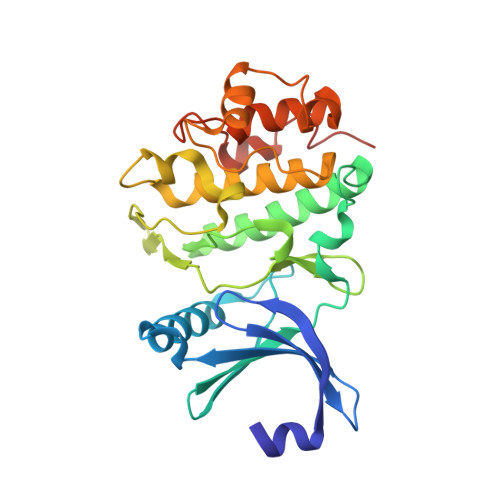
2YER

Synthesis and evaluation of triazolones as checkpoint kinase 1 inhibitors


Experimental Data Snapshot

  • Method: X-RAY DIFFRACTION
  • Resolution: 1.83 Å
  • R-Value Free: 0.175 
  • R-Value Work: 0.145 
  • R-Value Observed: 0.147 

wwPDB Validation   3D Report Full Report


Ligand Structure Quality Assessment 


This is version 1.1 of the entry. See complete history


Literature

Synthesis and Evaluation of Triazolones as Checkpoint Kinase 1 Inhibitors.

Oza, V.Ashwell, S.Brassil, P.Breed, J.Ezhuthachan, J.Deng, C.Grondine, M.Horn, C.Liu, D.Lyne, P.Newcombe, N.Pass, M.Read, J.Su, M.Toader, D.Yu, D.Yu, Y.Zabludoff, S.

(2012) Bioorg Med Chem Lett 22: 2330

  • DOI: https://doi.org/10.1016/j.bmcl.2012.01.043
  • Primary Citation of Related Structures:  
    2YER, 2YEX

  • PubMed Abstract: 

    Checkpoint kinase 1 (Chk1, CHEK1) is a Ser/Thr protein kinase that plays a key role in mediating the cellular response to DNA-damage. Synthesis and evaluation of a previously described class of Chk1 inhibitors, triazoloquinolones/triazolones (TZs) is further described herein. Our investigation of structure-activity relationships led to the identification of potent inhibitors 14c, 14h and 16e. Key challenges included modulation of physicochemical properties and pharmacokinetic (PK) parameters to enable compound testing in a Chk1 specific hollow fiber pharmacodynamic model. In this model, 16e was shown to abrogate topotecan-induced cell cycle arrest in a dose dependent manner. The demonstrated activity of TZs in this model in combination with a chemotherapeutic agent as well as radiotherapy validates this series of Chk1 inhibitors. X-ray crystal structures (PDB code: 2YEX and 2YER) for an initial lead and an optimized analog are also presented.


  • Organizational Affiliation

    AstraZeneca R&D Boston, 35 Gatehouse Drive, Waltham, MA 02451, USA. Vibha.Oza@astrazeneca.com


Macromolecules
Find similar proteins by:  (by identity cutoff)  |  3D Structure
Entity ID: 1
MoleculeChains Sequence LengthOrganismDetailsImage
SERINE/THREONINE-PROTEIN KINASE CHK1276Homo sapiensMutation(s): 0 
EC: 2.7.11.1
UniProt & NIH Common Fund Data Resources
Find proteins for O14757 (Homo sapiens)
Explore O14757 
Go to UniProtKB:  O14757
PHAROS:  O14757
GTEx:  ENSG00000149554 
Entity Groups  
Sequence Clusters30% Identity50% Identity70% Identity90% Identity95% Identity100% Identity
UniProt GroupO14757
Sequence Annotations
Expand
  • Reference Sequence
Small Molecules
Ligands 3 Unique
IDChains Name / Formula / InChI Key2D Diagram3D Interactions
TQ1
Query on TQ1

Download Ideal Coordinates CCD File 
B [auth A]5-(HYDROXYMETHYL)-8-(1H-PYRROL-2-YL)-2H-[1,2,4]TRIAZOLO[4,3-A]QUINOLIN-1-ONE
C15 H12 N4 O2
FEKDFTQZRBQDGS-UHFFFAOYSA-N
SO4
Query on SO4

Download Ideal Coordinates CCD File 
C [auth A]SULFATE ION
O4 S
QAOWNCQODCNURD-UHFFFAOYSA-L
EDO
Query on EDO

Download Ideal Coordinates CCD File 
D [auth A]
E [auth A]
F [auth A]
G [auth A]
H [auth A]
D [auth A],
E [auth A],
F [auth A],
G [auth A],
H [auth A],
I [auth A]
1,2-ETHANEDIOL
C2 H6 O2
LYCAIKOWRPUZTN-UHFFFAOYSA-N
Binding Affinity Annotations 
IDSourceBinding Affinity
TQ1 BindingDB:  2YER IC50: 20 (nM) from 1 assay(s)
PDBBind:  2YER IC50: 20 (nM) from 1 assay(s)
Binding MOAD:  2YER IC50: 20 (nM) from 1 assay(s)
Experimental Data & Validation

Experimental Data

  • Method: X-RAY DIFFRACTION
  • Resolution: 1.83 Å
  • R-Value Free: 0.175 
  • R-Value Work: 0.145 
  • R-Value Observed: 0.147 
  • Space Group: P 21 21 21
Unit Cell:
Length ( Å )Angle ( ˚ )
a = 41.16α = 90
b = 69.92β = 90
c = 103.5γ = 90
Software Package:
Software NamePurpose
REFMACrefinement
MOSFLMdata reduction
SCALAdata scaling
AMoREphasing

Structure Validation

View Full Validation Report


Ligand Structure Quality Assessment 


Entry History 

Deposition Data

Revision History  (Full details and data files)

  • Version 1.0: 2012-03-14
    Type: Initial release
  • Version 1.1: 2018-04-04
    Changes: Data collection