Discovery and Characterization of Small Molecule Inhibitors of the Bet Family Bromodomains.
Chung, C.W., Coste, H., White, J.H., Mirguet, O., Wilde, J., Gosmini, R.L., Delves, C., Magny, S.M., Woodward, R., Hughes, S.A., Boursier, E.V., Flynn, H., Bouillot, A.M., Bamborough, P., Brusq, J.M., Gellibert, F.J., Jones, E.J., Riou, A.M., Homes, P., Martin, S.L., Uings, I.J., Toum, J., Clement, C.A., Boullay, A.B., Grimley, R.L., Blandel, F.M., Prinjha, R.K., Lee, K., Kirilovsky, J., Nicodeme, E.(2011) J Med Chem 54: 3827
- PubMed: 21568322
- DOI: https://doi.org/10.1021/jm200108t
- Primary Citation of Related Structures:
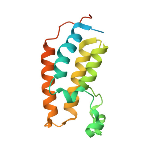
2YDW, 2YEK, 2YEL, 2YEM - PubMed Abstract:
Epigenetic mechanisms of gene regulation have a profound role in normal development and disease processes. An integral part of this mechanism occurs through lysine acetylation of histone tails which are recognized by bromodomains. While the biological and structural characterization of many bromodomain containing proteins has advanced considerably, the therapeutic tractability of this protein family is only now becoming understood. This paper describes the discovery and molecular characterization of potent (nM) small molecule inhibitors that disrupt the function of the BET family of bromodomains (Brd2, Brd3, and Brd4). By using a combination of phenotypic screening, chemoproteomics, and biophysical studies, we have discovered that the protein-protein interactions between bromodomains and acetylated histones can be antagonized by selective small molecules that bind at the acetylated lysine recognition pocket. X-ray crystal structures of compounds bound into bromodomains of Brd2 and Brd4 elucidate the molecular interactions of binding and explain the precisely defined stereochemistry required for activity.
Organizational Affiliation:
Molecular Discovery Research, GlaxoSmithKline R&D, Stevenage SG5 4HT, UK. Chun-Wa.H.Chung@gsk.com