2YB5

Structure of the fusidic acid resistance protein FusC


Experimental Data Snapshot

  • Method: X-RAY DIFFRACTION
  • Resolution: 2.10 Å
  • R-Value Free: 0.288 
  • R-Value Work: 0.213 
  • R-Value Observed: 0.217 

wwPDB Validation   3D Report Full Report


This is version 1.2 of the entry. See complete history


Literature

Ribosome Clearance by Fusb-Type Proteins Mediates Resistance to the Antibiotic Fusidic Acid

Cox, G.Thompson, G.S.Jenkins, H.T.Homans, S.W.Edwards, T.A.Oneill, A.J.

(2012) Proc Natl Acad Sci U S A 109: 2102

  • DOI: https://doi.org/10.1073/pnas.1117275109
  • Primary Citation of Related Structures:  
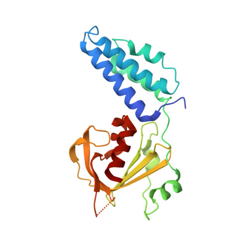
    2YB5

  • PubMed Abstract: 

    Resistance to the antibiotic fusidic acid (FA) in the human pathogen Staphylococcus aureus usually results from expression of FusB-type proteins (FusB or FusC). These proteins bind to elongation factor G (EF-G), the target of FA, and rescue translation from FA-mediated inhibition by an unknown mechanism. Here we show that the FusB family are two-domain metalloproteins, the C-terminal domain of which contains a four-cysteine zinc finger with a unique structural fold. This domain mediates a high-affinity interaction with the C-terminal domains of EF-G. By binding to EF-G on the ribosome, FusB-type proteins promote the dissociation of stalled ribosome⋅EF-G⋅GDP complexes that form in the presence of FA, thereby allowing the ribosomes to resume translation. Ribosome clearance by these proteins represents a highly unusual antibiotic resistance mechanism, which appears to be fine-tuned by the relative abundance of FusB-type protein, ribosomes, and EF-G.


  • Organizational Affiliation

    Institute of Molecular and Cellular Biology, University of Leeds, Leeds LS2 9JT, United Kingdom.


Macromolecules
Find similar proteins by:  (by identity cutoff)  |  3D Structure
Entity ID: 1
MoleculeChains Sequence LengthOrganismDetailsImage
PUTATIVE FUSIDIC ACID RESISTANCE PROTEINA,
B [auth F]
215Staphylococcus aureusMutation(s): 0 
UniProt
Find proteins for M1XHT1 (Staphylococcus aureus)
Explore M1XHT1 
Go to UniProtKB:  M1XHT1
Entity Groups  
Sequence Clusters30% Identity50% Identity70% Identity90% Identity95% Identity100% Identity
UniProt GroupM1XHT1
Sequence Annotations
Expand
  • Reference Sequence
Small Molecules
Ligands 2 Unique
IDChains Name / Formula / InChI Key2D Diagram3D Interactions
ZN
Query on ZN

Download Ideal Coordinates CCD File 
C [auth A],
K [auth F]
ZINC ION
Zn
PTFCDOFLOPIGGS-UHFFFAOYSA-N
EDO
Query on EDO

Download Ideal Coordinates CCD File 
D [auth F]
E [auth F]
F
G [auth F]
H [auth F]
D [auth F],
E [auth F],
F,
G [auth F],
H [auth F],
I [auth F],
J [auth F],
L [auth F]
1,2-ETHANEDIOL
C2 H6 O2
LYCAIKOWRPUZTN-UHFFFAOYSA-N
Experimental Data & Validation

Experimental Data

  • Method: X-RAY DIFFRACTION
  • Resolution: 2.10 Å
  • R-Value Free: 0.288 
  • R-Value Work: 0.213 
  • R-Value Observed: 0.217 
  • Space Group: P 1 21 1
Unit Cell:
Length ( Å )Angle ( ˚ )
a = 34.15α = 90
b = 109.61β = 101.72
c = 61.15γ = 90
Software Package:
Software NamePurpose
REFMACrefinement
MOSFLMdata reduction
SCALAdata scaling
SHELXphasing

Structure Validation

View Full Validation Report


Entry History 

Deposition Data

Revision History  (Full details and data files)

  • Version 1.0: 2012-01-25
    Type: Initial release
  • Version 1.1: 2012-02-29
    Changes: Other
  • Version 1.2: 2015-09-23
    Changes: Data collection