2Y7Q

THE HIGH-AFFINITY COMPLEX BETWEEN IGE AND ITS RECEPTOR FC EPSILON RI


Experimental Data Snapshot

  • Method: X-RAY DIFFRACTION
  • Resolution: 3.40 Å
  • R-Value Free: 0.292 
  • R-Value Work: 0.245 
  • R-Value Observed: 0.248 

wwPDB Validation   3D Report Full Report


Ligand Structure Quality Assessment 


This is version 2.1 of the entry. See complete history


Literature

Conformational Changes in Ige Contribute to its Uniquely Slow Dissociation Rate from Receptor Fceri

Holdom, M.D.Davies, A.M.Nettleship, J.E.Bagby, S.C.Dhaliwal, B.Girardi, E.Hunt, J.Gould, H.J.Beavil, A.J.Mcdonnell, J.M.Owens, R.J.Sutton, B.J.

(2011) Nat Struct Mol Biol 18: 571

  • DOI: https://doi.org/10.1038/nsmb.2044
  • Primary Citation of Related Structures:  
    2WQR, 2Y7Q

  • PubMed Abstract: 

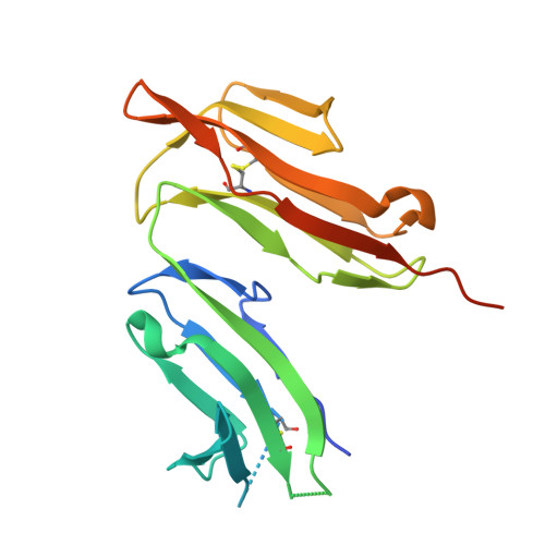
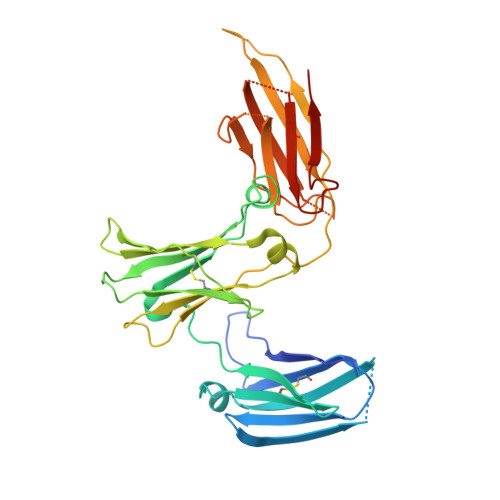
    Among antibody classes, IgE has a uniquely slow dissociation rate from, and high affinity for, its cell surface receptor FcɛRI. We show the structural basis for these key determinants of the ability of IgE to mediate allergic hypersensitivity through the 3.4-Å-resolution crystal structure of human IgE-Fc (consisting of the Cɛ2, Cɛ3 and Cɛ4 domains) bound to the extracellular domains of the FcɛRI α chain. Comparison with the structure of free IgE-Fc (reported here at a resolution of 1.9 Å) shows that the antibody, which has a compact, bent structure before receptor engagement, becomes even more acutely bent in the complex. Thermodynamic analysis indicates that the interaction is entropically driven, which explains how the noncontacting Cɛ2 domains, in place of the flexible hinge region of IgG antibodies, contribute together with the conformational changes to the unique binding properties of IgE.


  • Organizational Affiliation

    King's College London, Randall Division of Cell and Molecular Biophysics, London, UK.


Macromolecules
Find similar proteins by:  (by identity cutoff)  |  3D Structure
Entity ID: 1
MoleculeChains Sequence LengthOrganismDetailsImage
HIGH AFFINITY IMMUNOGLOBULIN EPSILON RECEPTOR SUBUNIT ALPHA188Homo sapiensMutation(s): 3 
UniProt & NIH Common Fund Data Resources
Find proteins for P12319 (Homo sapiens)
Explore P12319 
Go to UniProtKB:  P12319
PHAROS:  P12319
GTEx:  ENSG00000179639 
Entity Groups  
Sequence Clusters30% Identity50% Identity70% Identity90% Identity95% Identity100% Identity
UniProt GroupP12319
Sequence Annotations
Expand
  • Reference Sequence
Find similar proteins by:  (by identity cutoff)  |  3D Structure
Entity ID: 2
MoleculeChains Sequence LengthOrganismDetailsImage
IG EPSILON CHAIN C REGIONB,
C [auth D]
327Homo sapiensMutation(s): 3 
UniProt & NIH Common Fund Data Resources
Find proteins for P01854 (Homo sapiens)
Explore P01854 
Go to UniProtKB:  P01854
PHAROS:  P01854
GTEx:  ENSG00000211891 
Entity Groups  
Sequence Clusters30% Identity50% Identity70% Identity90% Identity95% Identity100% Identity
UniProt GroupP01854
Sequence Annotations
Expand
  • Reference Sequence
Oligosaccharides

Help

Entity ID: 3
MoleculeChains Length2D Diagram Glycosylation3D Interactions
alpha-D-mannopyranose-(1-3)-beta-D-mannopyranose-(1-4)-2-acetamido-2-deoxy-beta-D-glucopyranose-(1-4)-2-acetamido-2-deoxy-beta-D-glucopyranoseD [auth C]4N-Glycosylation
Glycosylation Resources
GlyTouCan:  G81315DD
GlyCosmos:  G81315DD
GlyGen:  G81315DD
Experimental Data & Validation

Experimental Data

  • Method: X-RAY DIFFRACTION
  • Resolution: 3.40 Å
  • R-Value Free: 0.292 
  • R-Value Work: 0.245 
  • R-Value Observed: 0.248 
  • Space Group: P 21 21 21
Unit Cell:
Length ( Å )Angle ( ˚ )
a = 99.491α = 90
b = 103.341β = 90
c = 110.122γ = 90
Software Package:
Software NamePurpose
PHENIXrefinement
MOSFLMdata reduction
SCALAdata scaling
MOLREPphasing

Structure Validation

View Full Validation Report


Ligand Structure Quality Assessment 


Entry History 

Deposition Data

Revision History  (Full details and data files)

  • Version 1.0: 2011-04-20
    Type: Initial release
  • Version 1.1: 2011-05-12
    Changes: Version format compliance
  • Version 1.2: 2011-07-13
    Changes: Version format compliance
  • Version 1.3: 2019-05-08
    Changes: Data collection, Experimental preparation, Other
  • Version 2.0: 2020-07-29
    Type: Remediation
    Reason: Carbohydrate remediation
    Changes: Atomic model, Data collection, Derived calculations, Other, Structure summary
  • Version 2.1: 2023-12-20
    Changes: Data collection, Database references, Derived calculations, Refinement description, Structure summary