2Y79

STRUCTURE OF THE FIRST GAF DOMAIN E87A MUTANT OF MYCOBACTERIUM TUBERCULOSIS DOSS


Experimental Data Snapshot

  • Method: X-RAY DIFFRACTION
  • Resolution: 1.80 Å
  • R-Value Free: 0.248 
  • R-Value Work: 0.197 
  • R-Value Observed: 0.199 

wwPDB Validation   3D Report Full Report


Ligand Structure Quality Assessment 


This is version 1.3 of the entry. See complete history


Literature

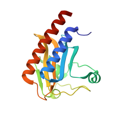
Blockage of the Channel to Heme by the E87 Side Chain in the Gaf Domain of Mycobacterium Tuberculosis Doss Confers the Unique Sensitivity of Doss to Oxygen.

Cho, H.Y.Cho, H.J.Kim, M.H.Kang, B.S.

(2011) FEBS Lett 585: 1873

  • DOI: https://doi.org/10.1016/j.febslet.2011.04.050
  • Primary Citation of Related Structures:  
    2Y79, 2Y8H

  • PubMed Abstract: 

    Two sensor kinases, DosS and DosT, are responsible for recognition of hypoxia in Mycobacterium tuberculosis. Both proteins are structurally similar to each other, but DosS is a redox sensor while DosT binds oxygen. The primary difference between the two proteins is the channel to the heme present in their GAF domains. DosS has a channel that is blocked by E87 while DosT has an open channel. Absorption spectra of DosS mutants with an open channel show that they bind oxygen as DosT does when they are exposed to air, while DosT G85E mutant is oxidized similarly to DosS without formation of an oxy-ferrous form. This suggests that oxygen accessibility to heme is the primary factor governing the oxygen-binding properties of these proteins.


  • Organizational Affiliation

    School of Life Science and Biotechnology, Kyungpook National University, Daegu, Republic of Korea.


Macromolecules
Find similar proteins by:  (by identity cutoff)  |  3D Structure
Entity ID: 1
MoleculeChains Sequence LengthOrganismDetailsImage
REDOX SENSOR HISTIDINE KINASE RESPONSE REGULATOR DEVS
A, B
153Mycobacterium tuberculosis H37RvMutation(s): 1 
EC: 2.7.13.3
UniProt
Find proteins for P9WGK3 (Mycobacterium tuberculosis (strain ATCC 25618 / H37Rv))
Explore P9WGK3 
Go to UniProtKB:  P9WGK3
Entity Groups  
Sequence Clusters30% Identity50% Identity70% Identity90% Identity95% Identity100% Identity
UniProt GroupP9WGK3
Sequence Annotations
Expand
  • Reference Sequence
Experimental Data & Validation

Experimental Data

  • Method: X-RAY DIFFRACTION
  • Resolution: 1.80 Å
  • R-Value Free: 0.248 
  • R-Value Work: 0.197 
  • R-Value Observed: 0.199 
  • Space Group: P 21 21 21
Unit Cell:
Length ( Å )Angle ( ˚ )
a = 35.932α = 90
b = 86.913β = 90
c = 100.973γ = 90
Software Package:
Software NamePurpose
REFMACrefinement
HKL-2000data scaling
AMoREphasing

Structure Validation

View Full Validation Report


Ligand Structure Quality Assessment 


Entry History 

Deposition Data

Revision History  (Full details and data files)

  • Version 1.0: 2011-02-16
    Type: Initial release
  • Version 1.1: 2011-06-16
    Changes: Version format compliance
  • Version 1.2: 2011-07-13
    Changes: Version format compliance
  • Version 1.3: 2023-12-20
    Changes: Data collection, Database references, Derived calculations, Other, Refinement description