2Y6B

Ascorbate Peroxidase R38K mutant


Experimental Data Snapshot

  • Method: X-RAY DIFFRACTION
  • Resolution: 1.90 Å
  • R-Value Free: 0.241 
  • R-Value Work: 0.175 
  • R-Value Observed: 0.178 

wwPDB Validation   3D Report Full Report


Ligand Structure Quality Assessment 


This is version 1.2 of the entry. See complete history


Literature

Proton Delivery to Ferryl Heme in a Heme Peroxidase: Enzymatic Use of the Grotthuss Mechanism.

Efimov, I.Badyal, S.K.Metcalfe, C.L.Macdonald, I.Gumiero, A.Raven, E.L.Moody, P.C.E.

(2011) J Am Chem Soc 133: 15376

  • DOI: https://doi.org/10.1021/ja2007017
  • Primary Citation of Related Structures:  
    2Y6A, 2Y6B

  • PubMed Abstract: 

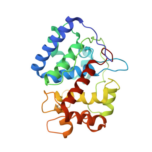
    We test the hypothesized pathway by which protons are passed from the substrate, ascorbate, to the ferryl oxygen in the heme enzyme ascorbate peroxidase (APX). The role of amino acid side chains and bound solvent is demonstrated. We investigated solvent kinetic isotope effects (SKIE) for the wild-type enzyme and several site-directed replacements of the key residues which form the proposed proton path. Kinetic constants for H(2)O(2)-dependent enzyme oxidation to Compound I, k(1), and subsequent reduction of Compound II, k(3), were determined in steady-state assays by variation of both H(2)O(2) and ascorbate concentrations. A high value of the SKIE for wild type APX ((D)k(3) = 4.9) as well as a clear nonlinear dependence on the deuterium composition of the solvent in proton inventory experiments suggest the simultaneous participation of several protons in the transition state for proton transfer. The full SKIE and the proton inventory data were modeled by applying Gross-Butler-Swain-Kresge theory to a proton path inferred from the known structure of APX. The model has been tested by constructing and determining the X-ray structures of the R38K and R38A variants and accounts for their observed SKIEs. This work confirms APX uses two arginine residues in the proton path. Thus, Arg38 and Arg172 have dual roles, both in the formation of the ferryl species and binding of ascorbate respectively and to facilitate proton transfer between the two.


  • Organizational Affiliation

    Department of Chemistry, Henry Wellcome Building, University of Leicester, University Road, Leicester LE1 7RH, England.


Macromolecules
Find similar proteins by:  (by identity cutoff)  |  3D Structure
Entity ID: 1
MoleculeChains Sequence LengthOrganismDetailsImage
ASCORBATE PEROXIDASE249Glycine maxMutation(s): 1 
EC: 1.11.1.11
UniProt
Find proteins for Q43758 (Glycine max)
Explore Q43758 
Go to UniProtKB:  Q43758
Entity Groups  
Sequence Clusters30% Identity50% Identity70% Identity90% Identity95% Identity100% Identity
UniProt GroupQ43758
Sequence Annotations
Expand
  • Reference Sequence
Small Molecules
Ligands 2 Unique
IDChains Name / Formula / InChI Key2D Diagram3D Interactions
HEM
Query on HEM

Download Ideal Coordinates CCD File 
B [auth A]PROTOPORPHYRIN IX CONTAINING FE
C34 H32 Fe N4 O4
KABFMIBPWCXCRK-RGGAHWMASA-L
SO4
Query on SO4

Download Ideal Coordinates CCD File 
C [auth A]SULFATE ION
O4 S
QAOWNCQODCNURD-UHFFFAOYSA-L
Experimental Data & Validation

Experimental Data

  • Method: X-RAY DIFFRACTION
  • Resolution: 1.90 Å
  • R-Value Free: 0.241 
  • R-Value Work: 0.175 
  • R-Value Observed: 0.178 
  • Space Group: P 42 21 2
Unit Cell:
Length ( Å )Angle ( ˚ )
a = 82.707α = 90
b = 82.707β = 90
c = 75.026γ = 90
Software Package:
Software NamePurpose
REFMACrefinement
iMOSFLMdata reduction
SCALAdata scaling
REFMACphasing

Structure Validation

View Full Validation Report


Ligand Structure Quality Assessment 


Entry History 

Deposition Data

Revision History  (Full details and data files)

  • Version 1.0: 2011-10-12
    Type: Initial release
  • Version 1.1: 2017-07-05
    Changes: Data collection
  • Version 1.2: 2023-12-20
    Changes: Data collection, Database references, Derived calculations, Other, Refinement description