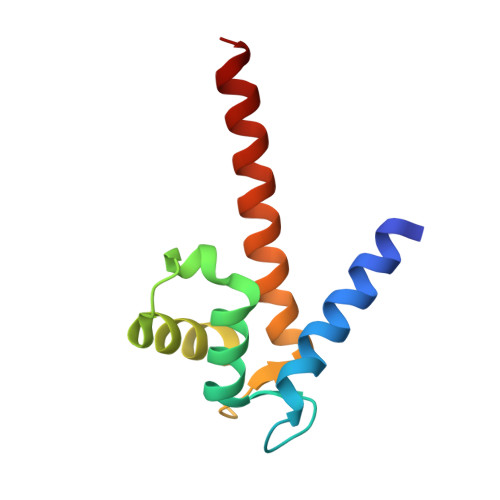
2Y5I

S100Z from zebrafish in complex with calcium


Experimental Data Snapshot

  • Method: X-RAY DIFFRACTION
  • Resolution: 2.03 Å
  • R-Value Free: 0.227 
  • R-Value Work: 0.171 
  • R-Value Observed: 0.174 

wwPDB Validation   3D Report Full Report


This is version 1.2 of the entry. See complete history


Literature

The Crystal Structure of Zebrafish S100Z: Implications for Calcium-Promoted S100 Protein Oligomerisation.

Moroz, O.V.Bronstein, I.B.Wilson, K.S.

(2011) J Mol Biol 411: 1072

  • DOI: https://doi.org/10.1016/j.jmb.2011.06.048
  • Primary Citation of Related Structures:  
    2Y5I

  • PubMed Abstract: 

    The S100 family, with about 20 members in humans, is composed of EF-hand calcium-regulated proteins and is linked to a range of serious human diseases, including cancer and autoimmune and neurological disorders. The oldest S100 family members are found in teleosts (bony fish). The zebrafish, Danio rerio, was suggested as a promising model system for in vivo studies on S100 family functions, and we chose to investigate zebrafish S100Z as the closest homologue of the metastasis-promoting human S100A4. Here, we report the first crystal structure of an S100 protein from this organism, the calcium-bound state of S100Z to 2.03 Å resolution. Crystal packing suggests higher-order oligomerisation of S100Z dimers, with a tetramerisation interface very similar to, but even more extensive than, that reported for S100A4. The interactions are primarily through the C-terminal αIV helices from adjacent dimers in an antiparallel orientation. Structural comparisons between known S100 multimeric assemblies together with analysis of calcium-driven changes to the dimerisation cores suggest a mechanism for calcium-promoted oligomerisation of S100 proteins.


  • Organizational Affiliation

    Structural Biology Laboratory, Department of Chemistry, University of York, Heslington, York YO10 5DD, UK. olga@ysbl.york.ac.uk


Macromolecules
Find similar proteins by:  (by identity cutoff)  |  3D Structure
Entity ID: 1
MoleculeChains Sequence LengthOrganismDetailsImage
S100 CALCIUM BINDING PROTEIN Z
A, B, C, D, E
A, B, C, D, E, F
99Danio rerioMutation(s): 0 
UniProt
Find proteins for Q503K9 (Danio rerio)
Explore Q503K9 
Go to UniProtKB:  Q503K9
Entity Groups  
Sequence Clusters30% Identity50% Identity70% Identity90% Identity95% Identity100% Identity
UniProt GroupQ503K9
Sequence Annotations
Expand
  • Reference Sequence
Small Molecules
Ligands 2 Unique
IDChains Name / Formula / InChI Key2D Diagram3D Interactions
IPA
Query on IPA

Download Ideal Coordinates CCD File 
I [auth A]
N [auth C]
Q [auth D]
T [auth E]
W [auth F]
I [auth A],
N [auth C],
Q [auth D],
T [auth E],
W [auth F],
X [auth F]
ISOPROPYL ALCOHOL
C3 H8 O
KFZMGEQAYNKOFK-UHFFFAOYSA-N
CA
Query on CA

Download Ideal Coordinates CCD File 
G [auth A]
H [auth A]
J [auth B]
K [auth B]
L [auth C]
G [auth A],
H [auth A],
J [auth B],
K [auth B],
L [auth C],
M [auth C],
O [auth D],
P [auth D],
R [auth E],
S [auth E],
U [auth F],
V [auth F]
CALCIUM ION
Ca
BHPQYMZQTOCNFJ-UHFFFAOYSA-N
Experimental Data & Validation

Experimental Data

  • Method: X-RAY DIFFRACTION
  • Resolution: 2.03 Å
  • R-Value Free: 0.227 
  • R-Value Work: 0.171 
  • R-Value Observed: 0.174 
  • Space Group: P 21 21 2
Unit Cell:
Length ( Å )Angle ( ˚ )
a = 75.611α = 90
b = 132.25β = 90
c = 58.209γ = 90
Software Package:
Software NamePurpose
REFMACrefinement
HKL-2000data reduction
SCALEPACKdata scaling
MOLREPphasing

Structure Validation

View Full Validation Report


Entry History 

Deposition Data

Revision History  (Full details and data files)

  • Version 1.0: 2011-07-27
    Type: Initial release
  • Version 1.1: 2011-08-24
    Changes: Database references
  • Version 1.2: 2023-12-20
    Changes: Data collection, Database references, Derived calculations, Other, Refinement description