2Y56

Fragment growing induces conformational changes in acetylcholine- binding protein: A structural and thermodynamic analysis - (Compound 3)


Experimental Data Snapshot

  • Method: X-RAY DIFFRACTION
  • Resolution: 3.59 Å
  • R-Value Free: 0.203 
  • R-Value Work: 0.174 
  • R-Value Observed: 0.175 

wwPDB Validation   3D Report Full Report


Ligand Structure Quality Assessment 


This is version 1.3 of the entry. See complete history


Literature

Fragment Growing Induces Conformational Changes in Acetylcholine-Binding Protein: A Structural and Thermodynamic Analysis.

Edink, E.Rucktooa, P.Retra, K.Akdemir, A.Nahar, T.Zuiderveld, O.Van Elk, R.Janssen, E.Van Nierop, P.Van Muijlwijk-Koezen, J.Smit, A.B.Sixma, T.K.Leurs, R.De Esch, I.J.P.

(2011) J Am Chem Soc 133: 5363

  • DOI: https://doi.org/10.1021/ja110571r
  • Primary Citation of Related Structures:  
    2Y54, 2Y56, 2Y57, 2Y58

  • PubMed Abstract: 

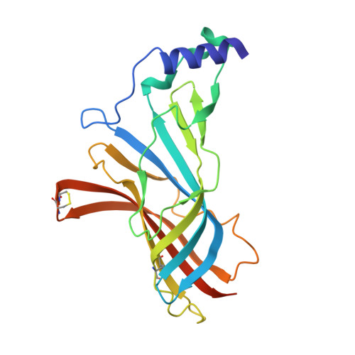
    Optimization of fragment hits toward high-affinity lead compounds is a crucial aspect of fragment-based drug discovery (FBDD). In the current study, we have successfully optimized a fragment by growing into a ligand-inducible subpocket of the binding site of acetylcholine-binding protein (AChBP). This protein is a soluble homologue of the ligand binding domain (LBD) of Cys-loop receptors. The fragment optimization was monitored with X-ray structures of ligand complexes and systematic thermodynamic analyses using surface plasmon resonance (SPR) biosensor analysis and isothermal titration calorimetry (ITC). Using site-directed mutagenesis and AChBP from different species, we find that specific changes in thermodynamic binding profiles, are indicative of interactions with the ligand-inducible subpocket of AChBP. This study illustrates that thermodynamic analysis provides valuable information on ligand binding modes and is complementary to affinity data when guiding rational structure- and fragment-based discovery approaches.


  • Organizational Affiliation

    Leiden/Amsterdam Center of Drug Research (LACDR), Division of Medicinal Chemistry, Faculty of Sciences, VU University Amsterdam, The Netherlands.


Macromolecules
Find similar proteins by:  (by identity cutoff)  |  3D Structure
Entity ID: 1
MoleculeChains Sequence LengthOrganismDetailsImage
SOLUBLE ACETYLCHOLINE RECEPTOR
A, B, C, D, E
217Aplysia californicaMutation(s): 0 
UniProt
Find proteins for Q8WSF8 (Aplysia californica)
Explore Q8WSF8 
Go to UniProtKB:  Q8WSF8
Entity Groups  
Sequence Clusters30% Identity50% Identity70% Identity90% Identity95% Identity100% Identity
UniProt GroupQ8WSF8
Sequence Annotations
Expand
  • Reference Sequence
Small Molecules
Ligands 4 Unique
IDChains Name / Formula / InChI Key2D Diagram3D Interactions
V11
Query on V11

Download Ideal Coordinates CCD File 
AA [auth D],
F [auth A],
KA [auth E],
M [auth B],
T [auth C]
[(1S,5R)-8-[(2R)-2-HYDROXY-2-PHENYL-ETHYL]-8-AZABICYCLO[3.2.1]OCTAN-3-YL] BENZOATE
C22 H25 N O3
CAELFFAMXVUOKR-BQJUDKOJSA-N
SO4
Query on SO4

Download Ideal Coordinates CCD File 
EA [auth D]
FA [auth D]
GA [auth D]
I [auth A]
J [auth A]
EA [auth D],
FA [auth D],
GA [auth D],
I [auth A],
J [auth A],
NA [auth E],
OA [auth E],
PA [auth E],
Q [auth B],
QA [auth E],
R [auth B],
RA [auth E],
W [auth C],
X [auth C]
SULFATE ION
O4 S
QAOWNCQODCNURD-UHFFFAOYSA-L
GOL
Query on GOL

Download Ideal Coordinates CCD File 
HA [auth D]
IA [auth D]
JA [auth D]
K [auth A]
L [auth A]
HA [auth D],
IA [auth D],
JA [auth D],
K [auth A],
L [auth A],
S [auth B],
Y [auth C],
Z [auth C]
GLYCEROL
C3 H8 O3
PEDCQBHIVMGVHV-UHFFFAOYSA-N
CL
Query on CL

Download Ideal Coordinates CCD File 
BA [auth D]
CA [auth D]
DA [auth D]
G [auth A]
H [auth A]
BA [auth D],
CA [auth D],
DA [auth D],
G [auth A],
H [auth A],
LA [auth E],
MA [auth E],
N [auth B],
O [auth B],
P [auth B],
U [auth C],
V [auth C]
CHLORIDE ION
Cl
VEXZGXHMUGYJMC-UHFFFAOYSA-M
Binding Affinity Annotations 
IDSourceBinding Affinity
V11 BindingDB:  2Y56 Ki: 100 (nM) from 1 assay(s)
PDBBind:  2Y56 Kd: 310 (nM) from 1 assay(s)
Experimental Data & Validation

Experimental Data

  • Method: X-RAY DIFFRACTION
  • Resolution: 3.59 Å
  • R-Value Free: 0.203 
  • R-Value Work: 0.174 
  • R-Value Observed: 0.175 
  • Space Group: I 2 3
Unit Cell:
Length ( Å )Angle ( ˚ )
a = 218.93α = 90
b = 218.93β = 90
c = 218.93γ = 90
Software Package:
Software NamePurpose
BUSTERrefinement
XDSdata reduction
XSCALEdata scaling
PHASERphasing

Structure Validation

View Full Validation Report


Ligand Structure Quality Assessment 


Entry History 

Deposition Data

Revision History  (Full details and data files)

  • Version 1.0: 2011-06-15
    Type: Initial release
  • Version 1.1: 2011-07-13
    Changes: Version format compliance
  • Version 1.2: 2019-04-24
    Changes: Data collection, Other, Source and taxonomy
  • Version 1.3: 2023-12-20
    Changes: Data collection, Database references, Derived calculations, Other, Refinement description