2Y3I

The structure of the fully closed conformation of human PGK in complex with L-ADP, 3PG and the TSA aluminium tetrafluoride


Experimental Data Snapshot

  • Method: X-RAY DIFFRACTION
  • Resolution: 2.90 Å
  • R-Value Free: 0.303 
  • R-Value Work: 0.263 
  • R-Value Observed: 0.265 

wwPDB Validation   3D Report Full Report


Ligand Structure Quality Assessment 


This is version 1.5 of the entry. See complete history


Literature

Interaction of Human 3-Phosphoglycerate Kinase with its Two Substrates: Is Substrate Antagonism a Kinetic Advantage?

Lallemand, P.Chaloin, L.Roy, B.Barman, T.Bowler, M.W.Lionne, C.

(2011) J Mol Biol 409: 742

  • DOI: https://doi.org/10.1016/j.jmb.2011.04.048
  • Primary Citation of Related Structures:  
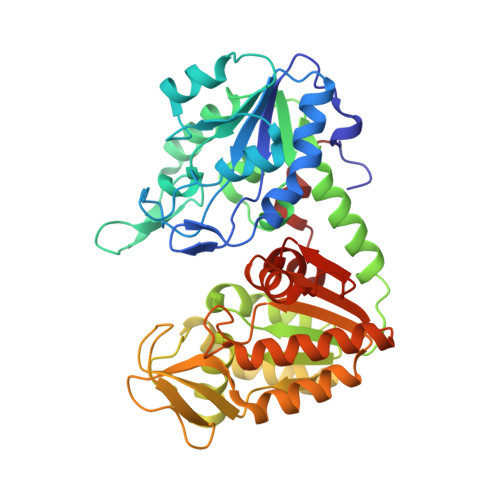
    2Y3I, 2YBE

  • PubMed Abstract: 

    Substrate antagonism has been described for a variety of enzymes with more than one substrate and is characterized by a lowering of the affinity of one substrate in the presence of the other(s). 3-Phosphoglycerate kinase (PGK) catalyzes phosphotransfer from 1,3-bisphosphoglycerate (bPG) to ADP to give 3-phosphoglycerate (PG) and ATP, and is subject to substrate antagonism. Because of the instability of bPG, antagonism has only been described between PG and ATP or ADP. Here, we show that antagonism also occurs between bPG and ADP. Using the stopped-flow method, we show that the dissociation constant for one substrate increases in the presence of the other, and that this decrease in affinity is mainly due to an increase in the dissociation rate constant. As a consequence, there is an increase in the overall interaction kinetics. Interestingly, in the presence of the mirror image of natural d-ADP, l-ADP (a good substrate for PGK), antagonism is absent. Using rapid-quench-flow, we studied the kinetics of ATP formation. The time courses present the following: (1) a lag with l-ADP, but not with d-ADP, the kinetics of which were similar to the interaction kinetics measured by stopped-flow; (2) a burst that is directed by the phosphotransfer; and (3) a steady-state that is rate limited by the release of product kinetics. Structural explanations for these results are proposed by analyzing the crystallographic structure of the fully closed conformation of PGK in complex with l-ADP, PG, and the transition-state analogue AlF(4)(-) compared to previously determined structures.


  • Organizational Affiliation

    Centre d'études d'agents Pathogènes et Biotechnologies pour le Santé, UMR 5236, CNRS-Universités Montpellier 1 and 2, 1919 route de Mende, 34293 Montpellier Cedex 5, France.


Macromolecules
Find similar proteins by:  (by identity cutoff)  |  3D Structure
Entity ID: 1
MoleculeChains Sequence LengthOrganismDetailsImage
PHOSPHOGLYCERATE KINASE 1A,
B [auth D]
416Homo sapiensMutation(s): 0 
EC: 2.7.2.3
UniProt & NIH Common Fund Data Resources
Find proteins for P00558 (Homo sapiens)
Explore P00558 
Go to UniProtKB:  P00558
PHAROS:  P00558
GTEx:  ENSG00000102144 
Entity Groups  
Sequence Clusters30% Identity50% Identity70% Identity90% Identity95% Identity100% Identity
UniProt GroupP00558
Sequence Annotations
Expand
  • Reference Sequence
Small Molecules
Ligands 5 Unique
IDChains Name / Formula / InChI Key2D Diagram3D Interactions
LA8
Query on LA8

Download Ideal Coordinates CCD File 
E [auth A],
H [auth D]
L-ADENOSINE-5'-DIPHOSPHATE
C10 H15 N5 O10 P2
XTWYTFMLZFPYCI-DEGSGYPDSA-N
3PG
Query on 3PG

Download Ideal Coordinates CCD File 
G [auth A],
L [auth D]
3-PHOSPHOGLYCERIC ACID
C3 H7 O7 P
OSJPPGNTCRNQQC-UWTATZPHSA-N
ALF
Query on ALF

Download Ideal Coordinates CCD File 
F [auth A],
I [auth D]
TETRAFLUOROALUMINATE ION
Al F4
UYOMQIYKOOHAMK-UHFFFAOYSA-J
CL
Query on CL

Download Ideal Coordinates CCD File 
D [auth A],
K [auth D]
CHLORIDE ION
Cl
VEXZGXHMUGYJMC-UHFFFAOYSA-M
MG
Query on MG

Download Ideal Coordinates CCD File 
C [auth A],
J [auth D]
MAGNESIUM ION
Mg
JLVVSXFLKOJNIY-UHFFFAOYSA-N
Binding Affinity Annotations 
IDSourceBinding Affinity
3PG BindingDB:  2Y3I Kd: 1.09e+5 (nM) from 1 assay(s)
Experimental Data & Validation

Experimental Data

  • Method: X-RAY DIFFRACTION
  • Resolution: 2.90 Å
  • R-Value Free: 0.303 
  • R-Value Work: 0.263 
  • R-Value Observed: 0.265 
  • Space Group: P 21 21 21
Unit Cell:
Length ( Å )Angle ( ˚ )
a = 38.419α = 90
b = 103.877β = 90
c = 203.094γ = 90
Software Package:
Software NamePurpose
REFMACrefinement
XDSdata reduction
SCALAdata scaling
MOLREPphasing

Structure Validation

View Full Validation Report


Ligand Structure Quality Assessment 


Entry History 

Deposition Data

Revision History  (Full details and data files)

  • Version 1.0: 2011-04-27
    Type: Initial release
  • Version 1.1: 2011-06-23
    Changes: Version format compliance
  • Version 1.2: 2011-07-13
    Changes: Version format compliance
  • Version 1.3: 2018-06-20
    Changes: Advisory, Data collection, Derived calculations
  • Version 1.4: 2019-01-23
    Changes: Advisory, Data collection, Derived calculations
  • Version 1.5: 2023-12-20
    Changes: Data collection, Database references, Derived calculations, Other, Refinement description