2Y38

LAMININ ALPHA5 CHAIN N-TERMINAL FRAGMENT


Experimental Data Snapshot

  • Method: X-RAY DIFFRACTION
  • Resolution: 2.90 Å
  • R-Value Free: 0.292 
  • R-Value Work: 0.254 
  • R-Value Observed: 0.254 

wwPDB Validation   3D Report Full Report


Ligand Structure Quality Assessment 


This is version 2.0 of the entry. See complete history


Literature

Determinants of Laminin Polymerisation Revealed by the Crystal Structure of the Alpha5 Chain Amino-Terminal Region

Hussain, S.A.Carafoli, F.Hohenester, E.

(2011) EMBO Rep 12: 276

  • DOI: https://doi.org/10.1038/embor.2011.3
  • Primary Citation of Related Structures:  
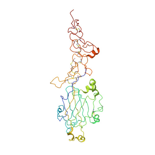
    2Y38

  • PubMed Abstract: 

    The polymerization of laminin into a cell-associated network--a key step in basement membrane assembly--is mediated by the laminin amino-terminal (LN) domains at the tips of the three short arms of the laminin αβγ-heterotrimer. The crystal structure of a laminin α5LN-LE1-2 fragment shows that the LN domain is a β-jelly roll with several elaborate insertions that is attached like a flower head to the stalk-like laminin-type epidermal growth factor-like tandem. A surface loop that is strictly conserved in the LN domains of all α-short arms is required for stable ternary association with the β- and γ-short arms in the laminin network.


  • Organizational Affiliation

    Department of Life Sciences, Biophysics Section, Blackett Laboratory, Prince Consort Road, Imperial College London, London SW7 2AZ, UK.


Macromolecules
Find similar proteins by:  (by identity cutoff)  |  3D Structure
Entity ID: 1
MoleculeChains Sequence LengthOrganismDetailsImage
LAMININ SUBUNIT ALPHA-5403Mus musculusMutation(s): 2 
UniProt & NIH Common Fund Data Resources
Find proteins for Q61001 (Mus musculus)
Explore Q61001 
Go to UniProtKB:  Q61001
IMPC:  MGI:105382
Entity Groups  
Sequence Clusters30% Identity50% Identity70% Identity90% Identity95% Identity100% Identity
UniProt GroupQ61001
Sequence Annotations
Expand
  • Reference Sequence
Oligosaccharides

Help

Entity ID: 2
MoleculeChains Length2D Diagram Glycosylation3D Interactions
2-acetamido-2-deoxy-beta-D-glucopyranose-(1-4)-2-acetamido-2-deoxy-beta-D-glucopyranose
B
2N-Glycosylation
Glycosylation Resources
GlyTouCan:  G42666HT
GlyCosmos:  G42666HT
GlyGen:  G42666HT
Experimental Data & Validation

Experimental Data

  • Method: X-RAY DIFFRACTION
  • Resolution: 2.90 Å
  • R-Value Free: 0.292 
  • R-Value Work: 0.254 
  • R-Value Observed: 0.254 
  • Space Group: P 62
Unit Cell:
Length ( Å )Angle ( ˚ )
a = 116.4α = 90
b = 116.4β = 90
c = 112.3γ = 120
Software Package:
Software NamePurpose
CNSrefinement
MOSFLMdata reduction
SCALAdata scaling
autoSHARPphasing

Structure Validation

View Full Validation Report


Ligand Structure Quality Assessment 


Entry History 

Deposition Data

Revision History  (Full details and data files)

  • Version 1.0: 2011-02-23
    Type: Initial release
  • Version 1.1: 2011-05-08
    Changes: Version format compliance
  • Version 1.2: 2011-07-13
    Changes: Version format compliance
  • Version 2.0: 2020-07-29
    Type: Remediation
    Reason: Carbohydrate remediation
    Changes: Atomic model, Data collection, Derived calculations, Other, Structure summary