2XZW

STRUCTURE OF PII FROM SYNECHOCOCCUS ELONGATUS IN COMPLEX WITH 2- OXOGLUTARATE AT LOW 2-OG CONCENTRATIONS


Experimental Data Snapshot

  • Method: X-RAY DIFFRACTION
  • Resolution: 1.95 Å
  • R-Value Free: 0.225 
  • R-Value Work: 0.175 
  • R-Value Observed: 0.178 

wwPDB Validation   3D Report Full Report


Ligand Structure Quality Assessment 


This is version 1.3 of the entry. See complete history


Literature

Mechanism of 2-Oxoglutarate Signaling by the Synechococcus Elongatus Pii Signal Transduction Protein.

Fokina, O.Chellamuthu, V.-R.Forchhammer, K.Zeth, K.

(2010) Proc Natl Acad Sci U S A 107: 19760

  • DOI: https://doi.org/10.1073/pnas.1007653107
  • Primary Citation of Related Structures:  
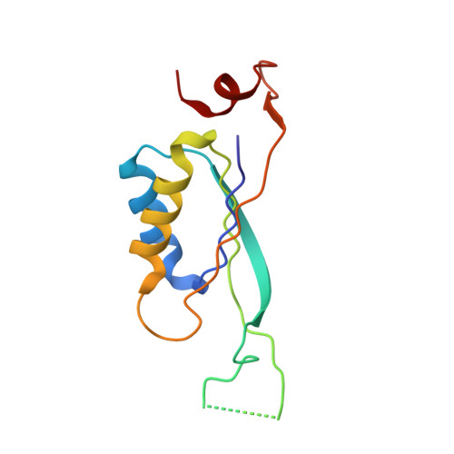
    2XUL, 2XZW

  • PubMed Abstract: 

    P(II) proteins control key processes of nitrogen metabolism in bacteria, archaea, and plants in response to the central metabolites ATP, ADP, and 2-oxoglutarate (2-OG), signaling cellular energy and carbon and nitrogen abundance. This metabolic information is integrated by P(II) and transmitted to regulatory targets (key enzymes, transporters, and transcription factors), modulating their activity. In oxygenic phototrophs, the controlling enzyme of arginine synthesis, N-acetyl-glutamate kinase (NAGK), is a major P(II) target, whose activity responds to 2-OG via P(II). Here we show structures of the Synechococcus elongatus P(II) protein in complex with ATP, Mg(2+), and 2-OG, which clarify how 2-OG affects P(II)-NAGK interaction. P(II) trimers with all three sites fully occupied were obtained as well as structures with one or two 2-OG molecules per P(II) trimer. These structures identify the site of 2-OG located in the vicinity between the subunit clefts and the base of the T loop. The 2-OG is bound to a Mg(2+) ion, which is coordinated by three phosphates of ATP, and by ionic interactions with the highly conserved residues K58 and Q39 together with B- and T-loop backbone interactions. These interactions impose a unique T-loop conformation that affects the interactions with the P(II) target. Structures of P(II) trimers with one or two bound 2-OG molecules reveal the basis for anticooperative 2-OG binding and shed light on the intersubunit signaling mechanism by which P(II) senses effectors in a wide range of concentrations.


  • Organizational Affiliation

    Interfakultäres Institut für Mikrobiologie und Infektionsmedizin der Eberhard-Karls-Universität Tübingen, Auf der Morgenstelle 28, 72076 Tübingen, Germany.


Macromolecules
Find similar proteins by:  (by identity cutoff)  |  3D Structure
Entity ID: 1
MoleculeChains Sequence LengthOrganismDetailsImage
NITROGEN REGULATORY PROTEIN P-II
A, B, C, D, E
A, B, C, D, E, F, G, H, I
115Synechococcus elongatus PCC 7942 = FACHB-805Mutation(s): 0 
UniProt
Find proteins for P0A3F4 (Synechococcus elongatus (strain ATCC 33912 / PCC 7942 / FACHB-805))
Explore P0A3F4 
Go to UniProtKB:  P0A3F4
Entity Groups  
Sequence Clusters30% Identity50% Identity70% Identity90% Identity95% Identity100% Identity
UniProt GroupP0A3F4
Sequence Annotations
Expand
  • Reference Sequence
Small Molecules
Ligands 3 Unique
IDChains Name / Formula / InChI Key2D Diagram3D Interactions
ATP
Query on ATP

Download Ideal Coordinates CCD File 
BA [auth I]
J [auth A]
M [auth B]
P [auth C]
S [auth D]
BA [auth I],
J [auth A],
M [auth B],
P [auth C],
S [auth D],
T [auth E],
U [auth F],
X [auth G],
Y [auth H]
ADENOSINE-5'-TRIPHOSPHATE
C10 H16 N5 O13 P3
ZKHQWZAMYRWXGA-KQYNXXCUSA-N
AKG
Query on AKG

Download Ideal Coordinates CCD File 
CA [auth I]
K [auth A]
N [auth B]
Q [auth C]
V [auth F]
CA [auth I],
K [auth A],
N [auth B],
Q [auth C],
V [auth F],
Z [auth H]
2-OXOGLUTARIC ACID
C5 H6 O5
KPGXRSRHYNQIFN-UHFFFAOYSA-N
MG
Query on MG

Download Ideal Coordinates CCD File 
AA [auth H]
DA [auth I]
L [auth A]
O [auth B]
R [auth C]
AA [auth H],
DA [auth I],
L [auth A],
O [auth B],
R [auth C],
W [auth F]
MAGNESIUM ION
Mg
JLVVSXFLKOJNIY-UHFFFAOYSA-N
Binding Affinity Annotations 
IDSourceBinding Affinity
AKG PDBBind:  2XZW Kd: 5100 (nM) from 1 assay(s)
Experimental Data & Validation

Experimental Data

  • Method: X-RAY DIFFRACTION
  • Resolution: 1.95 Å
  • R-Value Free: 0.225 
  • R-Value Work: 0.175 
  • R-Value Observed: 0.178 
  • Space Group: P 2 21 21
Unit Cell:
Length ( Å )Angle ( ˚ )
a = 72.334α = 90
b = 102.37β = 90
c = 135.774γ = 90
Software Package:
Software NamePurpose
REFMACrefinement
XDSdata reduction
XSCALEdata scaling
MOLREPphasing

Structure Validation

View Full Validation Report


Ligand Structure Quality Assessment 


Entry History 

Deposition Data

Revision History  (Full details and data files)

  • Version 1.0: 2010-12-08
    Type: Initial release
  • Version 1.1: 2011-05-08
    Changes: Version format compliance
  • Version 1.2: 2011-07-13
    Changes: Version format compliance
  • Version 1.3: 2023-12-20
    Changes: Data collection, Database references, Derived calculations, Other, Refinement description