2XZR

Escherichia coli Immunoglobulin-binding protein EibD 391-438 FUSED TO GCN4 ADAPTORS


Experimental Data Snapshot

  • Method: X-RAY DIFFRACTION
  • Resolution: 2.80 Å
  • R-Value Free: 0.308 
  • R-Value Work: 0.260 
  • R-Value Observed: 0.265 

wwPDB Validation   3D Report Full Report


This is version 1.1 of the entry. See complete history


Literature

The Structure of E. Coli Igg-Binding Protein D Suggests a General Model for Bending and Binding in Trimeric Autotransporter Adhesins.

Leo, J.C.Lyskowski, A.Hattula, K.Hartmann, M.D.Schwarz, H.Butcher, S.J.Linke, D.Lupas, A.N.Goldman, A.

(2011) Structure 19: 1021

  • DOI: https://doi.org/10.1016/j.str.2011.03.021
  • Primary Citation of Related Structures:  
    2XQH, 2XZR

  • PubMed Abstract: 

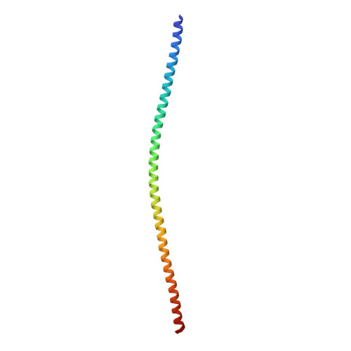
    The Escherichia coli Ig-binding (Eib) proteins are trimeric autotransporter adhesins (TAAs) and receptors for IgG Fc. We present the structure of a large fragment of the passenger domain of EibD, the first TAA structure to have both a YadA-like head domain and the entire coiled-coil stalk. The stalk begins as a right-handed superhelix, but switches handedness halfway down. An unexpected β-minidomain joins the two and inserts a ∼120° rotation such that there is no net twist between the beginning and end of the stalk. This may be important in folding and autotransport. The surprisingly large cavities we found in EibD and other TAAs may explain how TAAs bend to bind their ligands. We identified how IgA and IgG bind and modeled the EibD-IgG Fc complex. We further show that EibD promotes autoagglutination and biofilm formation and forms a fibrillar layer covering the cell surface making zipper-like contacts between cells.


  • Organizational Affiliation

    Macromolecular Crystallography Group, Institute of Biotechnology, University of Helsinki, 00014 Helsinki, Finland.


Macromolecules
Find similar proteins by:  (by identity cutoff)  |  3D Structure
Entity ID: 1
MoleculeChains Sequence LengthOrganismDetailsImage
IMMUNOGLOBULIN-BINDING PROTEIN EIBD114Enterobacteria phage P-EibDMutation(s): 0 
UniProt
Find proteins for Q9MCI8 (Escherichia coli)
Explore Q9MCI8 
Go to UniProtKB:  Q9MCI8
Entity Groups  
Sequence Clusters30% Identity50% Identity70% Identity90% Identity95% Identity100% Identity
UniProt GroupQ9MCI8
Sequence Annotations
Expand
  • Reference Sequence
Experimental Data & Validation

Experimental Data

  • Method: X-RAY DIFFRACTION
  • Resolution: 2.80 Å
  • R-Value Free: 0.308 
  • R-Value Work: 0.260 
  • R-Value Observed: 0.265 
  • Space Group: P 3 2 1
Unit Cell:
Length ( Å )Angle ( ˚ )
a = 36.67α = 90
b = 36.67β = 90
c = 228.58γ = 120
Software Package:
Software NamePurpose
REFMACrefinement
XDSdata reduction
XDSdata scaling
MOLREPphasing

Structure Validation

View Full Validation Report


Entry History 

Deposition Data

Revision History  (Full details and data files)

  • Version 1.0: 2011-07-20
    Type: Initial release
  • Version 1.1: 2023-12-20
    Changes: Data collection, Database references, Derived calculations, Other, Refinement description