2XYI

Crystal Structure of Nurf55 in complex with a H4 peptide


Experimental Data Snapshot

  • Method: X-RAY DIFFRACTION
  • Resolution: 1.75 Å
  • R-Value Free: 0.195 
  • R-Value Work: 0.161 
  • R-Value Observed: 0.162 

wwPDB Validation   3D Report Full Report


This is version 1.5 of the entry. See complete history


Literature

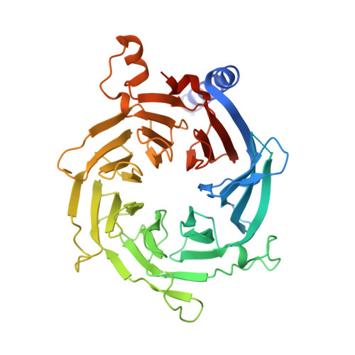
Chromatin-Modifying Complex Component Nurf55/P55 Associates with Histones H3, H4 and Polycomb Repressive Complex 2 Subunit Su(Z)12 Through Partially Overlapping Binding Sites.

Nowak, A.J.Alfieri, C.Stirnimann, C.U.Rybin, V.Baudin, F.Ly-Hartig, N.Lindner, D.Muller, C.W.

(2011) J Biol Chem 286: 23388

  • DOI: https://doi.org/10.1074/jbc.M110.207407
  • Primary Citation of Related Structures:  
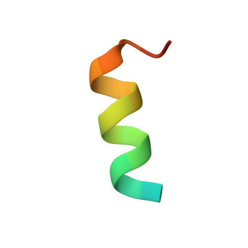
    2XYI

  • PubMed Abstract: 

    Drosophila Nurf55 is a component of different chromatin-modifying complexes, including the PRC2 (Polycomb repressive complex 2). Based on the 1.75-Å crystal structure of Nurf55 bound to histone H4 helix 1, we analyzed interactions of Nurf55 (Nurf55 or p55 in fly and RbAp48/46 in human) with the N-terminal tail of histone H3, the first helix of histone H4, and an N-terminal fragment of the PRC2 subunit Su(z)12 using isothermal calorimetry and pulldown experiments. Site-directed mutagenesis identified the binding site of histone H3 at the top of the Nurf55 WD40 propeller. Unmodified or K9me3- or K27me3-containing H3 peptides were bound with similar affinities, whereas the affinity for K4me3-containing H3 peptides was reduced. Helix 1 of histone H4 and Su(z)12 bound to the edge of the β-propeller using overlapping binding sites. Our results show similarities in the recognition of histone H4 and Su(z)12 and identify Nurf55 as a versatile interactor that simultaneously contacts multiple partners.


  • Organizational Affiliation

    European Molecular Biology Laboratory, 69117 Heidelberg, Germany.


Macromolecules
Find similar proteins by:  (by identity cutoff)  |  3D Structure
Entity ID: 1
MoleculeChains Sequence LengthOrganismDetailsImage
PROBABLE HISTONE-BINDING PROTEIN CAF1430Drosophila melanogasterMutation(s): 0 
UniProt
Find proteins for Q24572 (Drosophila melanogaster)
Explore Q24572 
Go to UniProtKB:  Q24572
Entity Groups  
Sequence Clusters30% Identity50% Identity70% Identity90% Identity95% Identity100% Identity
UniProt GroupQ24572
Sequence Annotations
Expand
  • Reference Sequence

Find similar proteins by:  Sequence   |   3D Structure  

Entity ID: 2
MoleculeChains Sequence LengthOrganismDetailsImage
HISTONE H420Drosophila melanogasterMutation(s): 0 
UniProt
Find proteins for P84040 (Drosophila melanogaster)
Explore P84040 
Go to UniProtKB:  P84040
Entity Groups  
Sequence Clusters30% Identity50% Identity70% Identity90% Identity95% Identity100% Identity
UniProt GroupP84040
Sequence Annotations
Expand
  • Reference Sequence
Experimental Data & Validation

Experimental Data

  • Method: X-RAY DIFFRACTION
  • Resolution: 1.75 Å
  • R-Value Free: 0.195 
  • R-Value Work: 0.161 
  • R-Value Observed: 0.162 
  • Space Group: P 1 21 1
Unit Cell:
Length ( Å )Angle ( ˚ )
a = 61.51α = 90
b = 59.21β = 99.83
c = 65.89γ = 90
Software Package:
Software NamePurpose
PHENIXrefinement
XDSdata reduction
XSCALEdata scaling
PHASERphasing

Structure Validation

View Full Validation Report


Entry History 

Deposition Data

Revision History  (Full details and data files)

  • Version 1.0: 2011-05-04
    Type: Initial release
  • Version 1.1: 2011-06-30
    Changes: Version format compliance
  • Version 1.2: 2011-07-13
    Changes: Version format compliance
  • Version 1.3: 2019-04-24
    Changes: Advisory, Data collection, Other, Source and taxonomy
  • Version 1.4: 2019-10-09
    Changes: Data collection, Experimental preparation, Other
  • Version 1.5: 2023-12-20
    Changes: Advisory, Data collection, Database references, Derived calculations, Refinement description