2XVY

Cobalt chelatase CbiK (periplasmic) from Desulvobrio vulgaris Hildenborough (co-crystallised with cobalt and SHC)


Experimental Data Snapshot

  • Method: X-RAY DIFFRACTION
  • Resolution: 1.70 Å
  • R-Value Free: 0.188 
  • R-Value Work: 0.172 
  • R-Value Observed: 0.173 

wwPDB Validation   3D Report Full Report


Ligand Structure Quality Assessment 


This is version 1.3 of the entry. See complete history


Literature

Desulfovibrio vulgaris CbiK(P) cobaltochelatase: evolution of a haem binding protein orchestrated by the incorporation of two histidine residues.

Lobo, S.A.Videira, M.A.Pacheco, I.Wass, M.N.Warren, M.J.Teixeira, M.Matias, P.M.Romao, C.V.Saraiva, L.M.

(2017) Environ Microbiol 19: 106-118

  • DOI: https://doi.org/10.1111/1462-2920.13479
  • Primary Citation of Related Structures:  
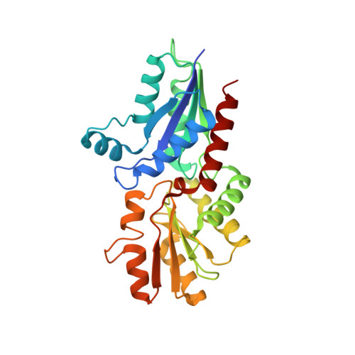
    2XVY

  • PubMed Abstract: 

    The sulfate-reducing bacteria of the Desulfovibrio genus make three distinct modified tetrapyrroles, haem, sirohaem and adenosylcobamide, where sirohydrochlorin acts as the last common biosynthetic intermediate along the branched tetrapyrrole pathway. Intriguingly, D. vulgaris encodes two sirohydrochlorin chelatases, CbiK P and CbiK C , that insert cobalt/iron into the tetrapyrrole macrocycle but are thought to be distinctly located in the periplasm and cytoplasm respectively. Fusing GFP onto the C-terminus of CbiK P confirmed that the protein is transported to the periplasm. The structure-function relationship of CbiK P was studied by constructing eleven site-directed mutants and determining their chelatase activities, oligomeric status and haem binding abilities. Residues His154 and His216 were identified as essential for metal-chelation of sirohydrochlorin. The tetrameric form of the protein is stabilized by Arg54 and Glu76, which form hydrogen bonds between two subunits. His96 is responsible for the binding of two haem groups within the main central cavity of the tetramer. Unexpectedly, CbiK P is shown to bind two additional haem groups through interaction with His103. Thus, although still retaining cobaltochelatase activity, the presence of His96 and His103 in CbiK P , which are absent from all other known bacterial cobaltochelatases, has evolved CbiK P a new function as a haem binding protein permitting it to act as a potential haem chaperone or transporter.


  • Organizational Affiliation

    Instituto de Tecnologia Química e Biológica NOVA, Avenida da República (EAN), Oeiras, 2780-157, Portugal.


Macromolecules
Find similar proteins by:  (by identity cutoff)  |  3D Structure
Entity ID: 1
MoleculeChains Sequence LengthOrganismDetailsImage
CHELATASE, PUTATIVE269Nitratidesulfovibrio vulgaris str. HildenboroughMutation(s): 0 
EC: 4.99.1.3
UniProt
Find proteins for Q72EC8 (Desulfovibrio vulgaris (strain ATCC 29579 / DSM 644 / NCIMB 8303 / VKM B-1760 / Hildenborough))
Explore Q72EC8 
Go to UniProtKB:  Q72EC8
Entity Groups  
Sequence Clusters30% Identity50% Identity70% Identity90% Identity95% Identity100% Identity
UniProt GroupQ72EC8
Sequence Annotations
Expand
  • Reference Sequence
Small Molecules
Ligands 6 Unique
IDChains Name / Formula / InChI Key2D Diagram3D Interactions
HEM
Query on HEM

Download Ideal Coordinates CCD File 
B [auth A]PROTOPORPHYRIN IX CONTAINING FE
C34 H32 Fe N4 O4
KABFMIBPWCXCRK-RGGAHWMASA-L
SO4
Query on SO4

Download Ideal Coordinates CCD File 
D [auth A]
E [auth A]
F [auth A]
G [auth A]
H [auth A]
D [auth A],
E [auth A],
F [auth A],
G [auth A],
H [auth A],
I [auth A]
SULFATE ION
O4 S
QAOWNCQODCNURD-UHFFFAOYSA-L
GOL
Query on GOL

Download Ideal Coordinates CCD File 
J [auth A]
K [auth A]
L [auth A]
M [auth A]
N [auth A]
J [auth A],
K [auth A],
L [auth A],
M [auth A],
N [auth A],
O [auth A],
P [auth A]
GLYCEROL
C3 H8 O3
PEDCQBHIVMGVHV-UHFFFAOYSA-N
CO
Query on CO

Download Ideal Coordinates CCD File 
C [auth A]COBALT (II) ION
Co
XLJKHNWPARRRJB-UHFFFAOYSA-N
PER
Query on PER

Download Ideal Coordinates CCD File 
Q [auth A]PEROXIDE ION
O2
ANAIPYUSIMHBEL-UHFFFAOYSA-N
NA
Query on NA

Download Ideal Coordinates CCD File 
R [auth A],
S [auth A]
SODIUM ION
Na
FKNQFGJONOIPTF-UHFFFAOYSA-N
Experimental Data & Validation

Experimental Data

  • Method: X-RAY DIFFRACTION
  • Resolution: 1.70 Å
  • R-Value Free: 0.188 
  • R-Value Work: 0.172 
  • R-Value Observed: 0.173 
  • Space Group: I 4 2 2
Unit Cell:
Length ( Å )Angle ( ˚ )
a = 120.81α = 90
b = 120.81β = 90
c = 119.71γ = 90
Software Package:
Software NamePurpose
REFMACrefinement
XDSdata reduction
SCALEPACKdata scaling
PHASERphasing

Structure Validation

View Full Validation Report


Ligand Structure Quality Assessment 


Entry History 

Deposition Data

Revision History  (Full details and data files)

  • Version 1.0: 2011-11-16
    Type: Initial release
  • Version 1.1: 2017-01-25
    Changes: Database references
  • Version 1.2: 2017-03-01
    Changes: Database references
  • Version 1.3: 2023-12-20
    Changes: Data collection, Database references, Derived calculations, Other, Refinement description