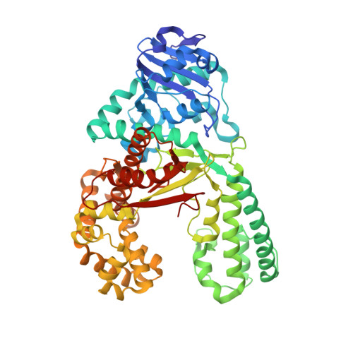
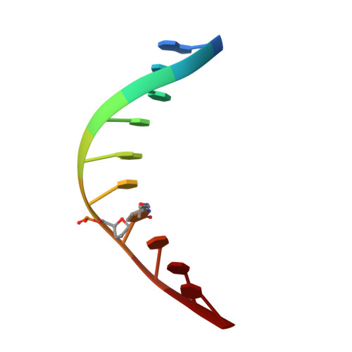
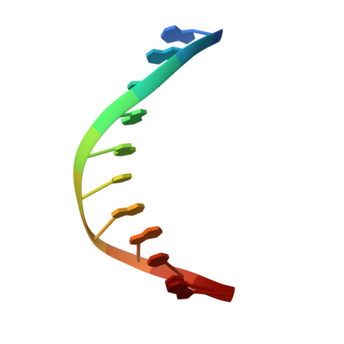
2XO7

Crystal structure of a dA:O-allylhydroxylamine-dC basepair in complex with fragment DNA polymerase I from Bacillus stearothermophilus


Experimental Data Snapshot

  • Method: X-RAY DIFFRACTION
  • Resolution: 2.85 Å
  • R-Value Free: 0.245 
  • R-Value Work: 0.215 
  • R-Value Observed: 0.216 

wwPDB Validation   3D Report Full Report


This is version 2.0 of the entry. See complete history


Literature

Chemical Discrimination between Dc and 5Medc Via Their Hydroxylamine Adducts

Muenzel, M.Lercher, L.Mueller, M.Carell, T.

(2010) Nucleic Acids Res 38: E192

  • DOI: https://doi.org/10.1093/nar/gkq724
  • Primary Citation of Related Structures:  
    2XO7

  • PubMed Abstract: 

    The presence of the methylated nucleobase (5Me)dC in CpG islands is a key factor that determines gene silencing. False methylation patterns are responsible for deteriorated cellular development and are a hallmark of many cancers. Today genes can be sequenced for the content of (5Me)dC only with the help of the bisulfite reagent, which is based exclusively on chemical reactivity differences established by the additional methyl group. Despite intensive optimization of the bisulfite protocol, the method still has specificity problems. Most importantly ∼95% of the DNA analyte is degraded during the analysis procedure. We discovered that the reagent O-allylhydroxylamine is able to discriminate between dC and (5Me)dC. The reagent, in contrast to bisulfite, does not exploit reactivity differences but gives directly different reaction products. The reagent forms a stable mutagenic adduct with dC, which can exist in two states (E versus Z). In case of dC the allylhydroxylamine adduct switches into the E-isomeric form, which generates dC to dT transition mutations that can easily be detected by established methods. Significantly, the (5Me)dC-adduct adopts exclusively the Z-isomeric form, which causes the polymerase to stop. O-allylhydroxylamine does allow differentiation between dC and (5Me)dC with high accuracy, leading towards a novel and mild chemistry for methylation analysis.


  • Organizational Affiliation

    Department of Chemistry, Center for Integrated Protein Science Munich, Ludwig-Maximilians University, Butenandtstr. 5-13, 81377 Munich, Germany.


Macromolecules

Find similar proteins by:  (by identity cutoff)  |  3D Structure
Entity ID: 1
MoleculeChains Sequence LengthOrganismDetailsImage
DNA POLYMERASE I580Geobacillus stearothermophilusMutation(s): 0 
EC: 2.7.7.7
UniProt
Find proteins for E1C9K5 (Geobacillus stearothermophilus)
Explore E1C9K5 
Go to UniProtKB:  E1C9K5
Entity Groups  
Sequence Clusters30% Identity50% Identity70% Identity90% Identity95% Identity100% Identity
UniProt GroupE1C9K5
Sequence Annotations
Expand
  • Reference Sequence

Find similar nucleic acids by:  Sequence   |   3D Structure  

Entity ID: 2
MoleculeChains LengthOrganismImage
5'-D(*GP*AP*CP*CP*AP*TP*47C*CP*CP*T)-3'10synthetic construct
Sequence Annotations
Expand
  • Reference Sequence

Find similar nucleic acids by:  Sequence   |   3D Structure  

Entity ID: 3
MoleculeChains LengthOrganismImage
5'-D(*AP*GP*GP*AP*AP*TP*GP*GP*TP*CP*A)-3'11synthetic construct
Sequence Annotations
Expand
  • Reference Sequence
Experimental Data & Validation

Experimental Data

  • Method: X-RAY DIFFRACTION
  • Resolution: 2.85 Å
  • R-Value Free: 0.245 
  • R-Value Work: 0.215 
  • R-Value Observed: 0.216 
  • Space Group: P 21 21 21
Unit Cell:
Length ( Å )Angle ( ˚ )
a = 87.478α = 90
b = 93.636β = 90
c = 105.091γ = 90
Software Package:
Software NamePurpose
REFMACrefinement
XDSdata reduction
SCALAdata scaling
PHASERphasing

Structure Validation

View Full Validation Report


Entry History 

Deposition Data

Revision History  (Full details and data files)

  • Version 1.0: 2010-09-15
    Type: Initial release
  • Version 1.1: 2011-09-21
    Changes: Database references, Version format compliance
  • Version 2.0: 2023-12-20
    Changes: Data collection, Database references, Derived calculations, Other, Polymer sequence, Refinement description