2XJA

Structure of MurE from M.tuberculosis with dipeptide and ADP


Experimental Data Snapshot

  • Method: X-RAY DIFFRACTION
  • Resolution: 3.00 Å
  • R-Value Free: 0.258 
  • R-Value Work: 0.189 
  • R-Value Observed: 0.193 

wwPDB Validation   3D Report Full Report


Ligand Structure Quality Assessment 


This is version 1.3 of the entry. See complete history


Literature

Essential Residues for the Enzyme Activity of ATP-Dependent Mure Ligase from Mycobacterium Tuberculosis.

Basavannacharya, C.Moody, P.R.Munshi, T.Cronin, N.Keep, N.H.Bhakta, S.

(2010) Protein Cell 1: 1011

  • DOI: https://doi.org/10.1007/s13238-010-0132-9
  • Primary Citation of Related Structures:  
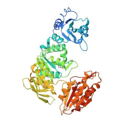
    2XJA

  • PubMed Abstract: 

    The emergence of total drug-resistant tuberculosis (TDRTB) has made the discovery of new therapies for tuberculosis urgent. The cytoplasmic enzymes of peptidoglycan biosynthesis have generated renewed interest as attractive targets for the development of new anti-mycobacterials. One of the cytoplasmic enzymes, uridine diphosphate (UDP)-MurNAc-tripeptide ligase (MurE), catalyses the addition of meso-diaminopimelic acid (m-DAP) into peptidoglycan in Mycobacterium tuberculosis coupled to the hydrolysis of ATP. Mutants of M. tuberculosis MurE were generated by replacing K157, E220, D392, R451 with alanine and N449 with aspartate, and truncating the first 24 amino acid residues at the N-terminus of the enzyme. Analysis of the specific activity of these proteins suggested that apart from the 24 N-terminal residues, the other mutated residues are essential for catalysis. Variations in K(m) values for one or more substrates were observed for all mutants, except the N-terminal truncation mutant, indicating that these residues are involved in binding substrates and form part of the active site structure. These mutant proteins were also tested for their specificity for a wide range of substrates. Interestingly, the mutations K157A, E220A and D392A showed hydrolysis of ATP uncoupled from catalysis. The ATP hydrolysis rate was enhanced by at least partial occupation of the uridine nucleotide dipeptide binding site. This study provides an insight into the residues essential for the catalytic activity and substrate binding of the ATP-dependent MurE ligase. Since ATP-dependent MurE ligase is a novel drug target, the understanding of its function may lead to development of novel inhibitors against resistant forms of M. tuberculosis.


  • Organizational Affiliation

    Department of Biological Sciences, Institute for Structural and Molecular Biology, Birkbeck, UK.


Macromolecules
Find similar proteins by:  (by identity cutoff)  |  3D Structure
Entity ID: 1
MoleculeChains Sequence LengthOrganismDetailsImage
UDP-N-ACETYLMURAMOYL-L-ALANYL-D-GLUTAMATE--2,6-DIAMINOPIMELATE LIGASE
A, B, C, D
535Mycobacterium tuberculosis H37RvMutation(s): 0 
EC: 6.3.2.13
UniProt
Find proteins for P9WJL3 (Mycobacterium tuberculosis (strain ATCC 25618 / H37Rv))
Explore P9WJL3 
Go to UniProtKB:  P9WJL3
Entity Groups  
Sequence Clusters30% Identity50% Identity70% Identity90% Identity95% Identity100% Identity
UniProt GroupP9WJL3
Sequence Annotations
Expand
  • Reference Sequence
Small Molecules
Ligands 3 Unique
IDChains Name / Formula / InChI Key2D Diagram3D Interactions
UAG
Query on UAG

Download Ideal Coordinates CCD File 
H [auth A],
L [auth B],
P [auth C],
U [auth D]
URIDINE-5'-DIPHOSPHATE-N-ACETYLMURAMOYL-L-ALANINE-D-GLUTAMATE
C28 H43 N5 O23 P2
OJZCATPXPWFLHF-HPUCEMLMSA-N
ADP
Query on ADP

Download Ideal Coordinates CCD File 
E [auth A],
J [auth B],
N [auth C],
S [auth D]
ADENOSINE-5'-DIPHOSPHATE
C10 H15 N5 O10 P2
XTWYTFMLZFPYCI-KQYNXXCUSA-N
MG
Query on MG

Download Ideal Coordinates CCD File 
F [auth A]
G [auth A]
I [auth B]
K [auth B]
M [auth B]
F [auth A],
G [auth A],
I [auth B],
K [auth B],
M [auth B],
O [auth C],
Q [auth C],
R [auth D],
T [auth D],
V [auth D]
MAGNESIUM ION
Mg
JLVVSXFLKOJNIY-UHFFFAOYSA-N
Modified Residues  1 Unique
IDChains TypeFormula2D DiagramParent
KCX
Query on KCX
A, B, C, D
L-PEPTIDE LINKINGC7 H14 N2 O4LYS
Experimental Data & Validation

Experimental Data

  • Method: X-RAY DIFFRACTION
  • Resolution: 3.00 Å
  • R-Value Free: 0.258 
  • R-Value Work: 0.189 
  • R-Value Observed: 0.193 
  • Space Group: P 1
Unit Cell:
Length ( Å )Angle ( ˚ )
a = 75.04α = 111.32
b = 76.32β = 91.42
c = 81.99γ = 92.9
Software Package:
Software NamePurpose
REFMACrefinement
MOSFLMdata reduction
SCALAdata scaling
SCALAphasing

Structure Validation

View Full Validation Report


Ligand Structure Quality Assessment 


Entry History 

Deposition Data

Revision History  (Full details and data files)

  • Version 1.0: 2010-08-18
    Type: Initial release
  • Version 1.1: 2011-05-08
    Changes: Version format compliance
  • Version 1.2: 2011-07-13
    Changes: Version format compliance
  • Version 1.3: 2023-12-20
    Changes: Data collection, Database references, Derived calculations, Other, Refinement description