2XHS

Crystal structure of the ligand binding domain of Fushi tarazu factor 1 of Drosophila melanogaster.


Experimental Data Snapshot

  • Method: X-RAY DIFFRACTION
  • Resolution: 2.80 Å
  • R-Value Free: 0.246 
  • R-Value Work: 0.217 
  • R-Value Observed: 0.219 

wwPDB Validation   3D Report Full Report


This is version 1.2 of the entry. See complete history


Literature

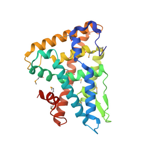
Crystal Structure of Fushi Tarazu Factor 1 Ligand Binding Domain/Fushi Tarazu Peptide Complex Identifies New Class of Nuclear Receptors.

Yoo, J.Ko, S.Kim, H.Sampson, H.Yun, J.H.Choe, K.M.Chang, I.Arrowsmith, C.H.Krause, H.M.Cho, H.S.Lee, W.

(2011) J Biol Chem 286: 31225

  • DOI: https://doi.org/10.1074/jbc.M111.252916
  • Primary Citation of Related Structures:  
    2XHS

  • PubMed Abstract: 

    The interaction between the orphan nuclear receptor FTZ-F1 (Fushi tarazu factor 1) and the segmentation gene protein FTZ is critical for specifying alternate parasegments in the Drosophila embryo. Here, we have determined the structure of the FTZ-F1 ligand-binding domain (LBD)·FTZ peptide complex using x-ray crystallography. Strikingly, the ligand-binding pocket of the FTZ-F1 LBD is completely occupied by helix 6 (H6) of the receptor, whereas the cofactor FTZ binds the co-activator cleft site of the FTZ-F1 LBD. Our findings suggest that H6 is essential for transcriptional activity of FTZ-F1; this is further supported by data from mutagenesis and activity assays. These data suggest that FTZ-F1 might belong to a novel class of ligand-independent nuclear receptors. Our findings are intriguing given that the highly homologous human steroidogenic factor-1 and liver receptor homolog-1 LBDs exhibit sizable ligand-binding pockets occupied by putative ligand molecules.


  • Organizational Affiliation

    Department of Biology, College of Life Science and Biotechnology, Yonsei University, Shinchon-dong, Seodaemun-gu 134, Seoul 120-749, Korea.


Macromolecules
Find similar proteins by:  (by identity cutoff)  |  3D Structure
Entity ID: 1
MoleculeChains Sequence LengthOrganismDetailsImage
NUCLEAR HORMONE RECEPTOR FTZ-F1245Drosophila melanogasterMutation(s): 0 
UniProt
Find proteins for P33244 (Drosophila melanogaster)
Explore P33244 
Go to UniProtKB:  P33244
Entity Groups  
Sequence Clusters30% Identity50% Identity70% Identity90% Identity95% Identity100% Identity
UniProt GroupP33244
Sequence Annotations
Expand
  • Reference Sequence

Find similar proteins by:  Sequence   |   3D Structure  

Entity ID: 2
MoleculeChains Sequence LengthOrganismDetailsImage
SEGMENTATION PROTEIN FUSHI TARAZU9Drosophila melanogasterMutation(s): 0 
UniProt
Find proteins for P02835 (Drosophila melanogaster)
Explore P02835 
Go to UniProtKB:  P02835
Entity Groups  
UniProt GroupP02835
Sequence Annotations
Expand
  • Reference Sequence
Small Molecules
Modified Residues  1 Unique
IDChains TypeFormula2D DiagramParent
MSE
Query on MSE
A
L-PEPTIDE LINKINGC5 H11 N O2 SeMET
Experimental Data & Validation

Experimental Data

  • Method: X-RAY DIFFRACTION
  • Resolution: 2.80 Å
  • R-Value Free: 0.246 
  • R-Value Work: 0.217 
  • R-Value Observed: 0.219 
  • Space Group: P 31 2 1
Unit Cell:
Length ( Å )Angle ( ˚ )
a = 97.931α = 90
b = 97.931β = 90
c = 123.466γ = 120
Software Package:
Software NamePurpose
REFMACrefinement
HKL-2000data reduction
SCALEPACKdata scaling
SOLVEphasing

Structure Validation

View Full Validation Report


Entry History 

Deposition Data

Revision History  (Full details and data files)

  • Version 1.0: 2011-07-20
    Type: Initial release
  • Version 1.1: 2011-09-14
    Changes: Database references
  • Version 1.2: 2018-06-20
    Changes: Advisory, Data collection, Derived calculations