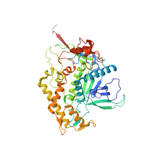
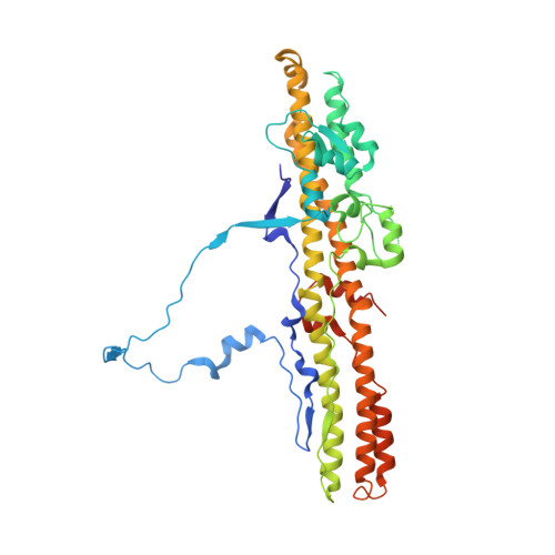
2XHL

Structure of a functional derivative of Clostridium botulinum neurotoxin type B


Experimental Data Snapshot

  • Method: X-RAY DIFFRACTION
  • Resolution: 2.80 Å
  • R-Value Free: 0.282 
  • R-Value Work: 0.240 
  • R-Value Observed: 0.242 

wwPDB Validation   3D Report Full Report


This is version 2.0 of the entry. See complete history


Literature

Structure and Activity of a Functional Derivative of Clostridium Botulinum Neurotoxin B.

Masuyer, G.Beard, M.Cadd, V.A.Chaddock, J.A.Acharya, K.R.

(2011) J Struct Biol 174: 52

  • DOI: https://doi.org/10.1016/j.jsb.2010.11.010
  • Primary Citation of Related Structures:  
    2XHL

  • PubMed Abstract: 

    Botulinum neurotoxins (BoNTs) cause flaccid paralysis by inhibiting neurotransmission at cholinergic nerve terminals. BoNTs consist of three essential domains for toxicity: the cell binding domain (Hc), the translocation domain (Hn) and the catalytic domain (LC). A functional derivative (LHn) of the parent neurotoxin B composed of Hn and LC domains was recombinantly produced and characterised. LHn/B crystallographic structure at 2.8Å resolution is reported. The catalytic activity of LHn/B towards recombinant human VAMP was analysed by substrate cleavage assay and showed a higher specificity for VAMP-1, -2 compared to VAMP-3. LHn/B also showed measurable activity in living spinal cord neurons. Despite lacking the Hc (cell-targeting) domain, LHn/B retained the capacity to internalize and cleave intracellular VAMP-1 and -2 when added to the cells at high concentration. These activities of the LHn/B fragment demonstrate the utility of engineered botulinum neurotoxin fragments as analytical tools to study the mechanisms of action of BoNT neurotoxins and of SNARE proteins.


  • Organizational Affiliation

    Department of Biology and Biochemistry, University of Bath, Claverton Down, Bath BA2 7AY, United Kingdom.


Macromolecules
Find similar proteins by:  (by identity cutoff)  |  3D Structure
Entity ID: 1
MoleculeChains Sequence LengthOrganismDetailsImage
BOTULINUM NEUROTOXIN B LIGHT CHAIN453Clostridium botulinumMutation(s): 0 
EC: 3.4.24.69
UniProt
Find proteins for P10844 (Clostridium botulinum)
Explore P10844 
Go to UniProtKB:  P10844
Entity Groups  
Sequence Clusters30% Identity50% Identity70% Identity90% Identity95% Identity100% Identity
UniProt GroupP10844
Sequence Annotations
Expand
  • Reference Sequence
Find similar proteins by:  (by identity cutoff)  |  3D Structure
Entity ID: 2
MoleculeChains Sequence LengthOrganismDetailsImage
BOTULINUM NEUROTOXIN B HEAVY CHAIN433Clostridium botulinumMutation(s): 0 
UniProt
Find proteins for P10844 (Clostridium botulinum)
Explore P10844 
Go to UniProtKB:  P10844
Entity Groups  
Sequence Clusters30% Identity50% Identity70% Identity90% Identity95% Identity100% Identity
UniProt GroupP10844
Sequence Annotations
Expand
  • Reference Sequence
Small Molecules
Ligands 1 Unique
IDChains Name / Formula / InChI Key2D Diagram3D Interactions
ZN
Query on ZN

Download Ideal Coordinates CCD File 
C [auth A]ZINC ION
Zn
PTFCDOFLOPIGGS-UHFFFAOYSA-N
Experimental Data & Validation

Experimental Data

  • Method: X-RAY DIFFRACTION
  • Resolution: 2.80 Å
  • R-Value Free: 0.282 
  • R-Value Work: 0.240 
  • R-Value Observed: 0.242 
  • Space Group: P 21 21 2
Unit Cell:
Length ( Å )Angle ( ˚ )
a = 66.89α = 90
b = 149.1β = 90
c = 113.49γ = 90
Software Package:
Software NamePurpose
REFMACrefinement
MOSFLMdata reduction
SCALAdata scaling
PHASERphasing

Structure Validation

View Full Validation Report


Entry History 

Deposition Data

Revision History  (Full details and data files)

  • Version 1.0: 2010-12-01
    Type: Initial release
  • Version 1.1: 2011-06-30
    Changes: Version format compliance
  • Version 1.2: 2011-07-13
    Changes: Version format compliance
  • Version 1.3: 2023-12-20
    Changes: Data collection, Database references, Derived calculations, Other, Refinement description
  • Version 2.0: 2024-01-31
    Changes: Atomic model