2XGW

ZINC-BOUND CRYSTAL STRUCTURE OF STREPTOCOCCUS PYOGENES DPR


Experimental Data Snapshot

  • Method: X-RAY DIFFRACTION
  • Resolution: 2.10 Å
  • R-Value Free: 0.172 
  • R-Value Work: 0.144 
  • R-Value Observed: 0.145 

wwPDB Validation   3D Report Full Report


This is version 1.3 of the entry. See complete history


Literature

Structural Characterization and Biological Implications of Di-Zinc Binding in the Ferroxidase Center of Streptococcus Pyogenes Dpr.

Haikarainen, T.Tsou, C.-C.Wu, J.-J.Papageorgiou, A.C.

(2010) Biochem Biophys Res Commun 398: 361

  • DOI: https://doi.org/10.1016/j.bbrc.2010.06.071
  • Primary Citation of Related Structures:  
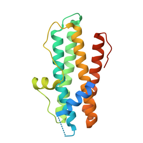
    2XGW

  • PubMed Abstract: 

    Dps proteins contain a ferroxidase site that binds and oxidizes iron, thereby preventing hydroxyl radical formation by Fenton reaction. Although the involvement of a di-iron ferroxidase site has been suggested, X-ray crystal structures of various Dps members have shown either one or two iron cations with various occupancies despite the high structural conservation of the site. Similarly, structural studies with zinc, a redox-stable replacement for iron, have shown the binding of either one or two zinc ions. Here, the crystal structure of Streptococcus pyogenes Dpr in complex with zinc reveals the binding of two zinc cations in the ferroxidase center and an additional zinc-binding site at the surface of the protein. The results suggest a structural basis for the protection of Streptococcus pyogenes in zinc stress conditions and provide a clear evidence for a di-zinc and di-iron ferroxidase site in Streptococcus pyogenes Dpr protein.


  • Organizational Affiliation

    Turku Centre for Biotechnology, University of Turku and Abo Akademi University, Finland.


Macromolecules
Find similar proteins by:  (by identity cutoff)  |  3D Structure
Entity ID: 1
MoleculeChains Sequence LengthOrganismDetailsImage
PEROXIDE RESISTANCE PROTEIN199Streptococcus pyogenesMutation(s): 0 
UniProt
Find proteins for Q99YU7 (Streptococcus pyogenes serotype M1)
Explore Q99YU7 
Go to UniProtKB:  Q99YU7
Entity Groups  
Sequence Clusters30% Identity50% Identity70% Identity90% Identity95% Identity100% Identity
UniProt GroupQ99YU7
Sequence Annotations
Expand
  • Reference Sequence
Experimental Data & Validation

Experimental Data

  • Method: X-RAY DIFFRACTION
  • Resolution: 2.10 Å
  • R-Value Free: 0.172 
  • R-Value Work: 0.144 
  • R-Value Observed: 0.145 
  • Space Group: F 41 3 2
Unit Cell:
Length ( Å )Angle ( ˚ )
a = 187.12α = 90
b = 187.12β = 90
c = 187.12γ = 90
Software Package:
Software NamePurpose
REFMACrefinement
MOSFLMdata reduction
SCALAdata scaling

Structure Validation

View Full Validation Report


Entry History 

Deposition Data

Revision History  (Full details and data files)

  • Version 1.0: 2010-08-11
    Type: Initial release
  • Version 1.1: 2011-05-08
    Changes: Version format compliance
  • Version 1.2: 2011-07-13
    Changes: Version format compliance
  • Version 1.3: 2019-07-24
    Changes: Data collection, Derived calculations