2XEU

Ring domain


Experimental Data Snapshot

  • Method: X-RAY DIFFRACTION
  • Resolution: 1.50 Å
  • R-Value Free: 0.197 
  • R-Value Work: 0.168 
  • R-Value Observed: 0.170 

wwPDB Validation   3D Report Full Report


This is version 2.0 of the entry. See complete history


Literature

Mechanism of Ubiquitylation by Dimeric Ring Ligase Rnf4

Plechanovova, A.Jaffray, E.G.Mcmahon, S.A.Johnson, K.A.Navratilova, I.Naismith, J.H.Hay, R.T.

(2011) Nat Struct Mol Biol 18: 1052

  • DOI: https://doi.org/10.1038/nsmb.2108
  • Primary Citation of Related Structures:  
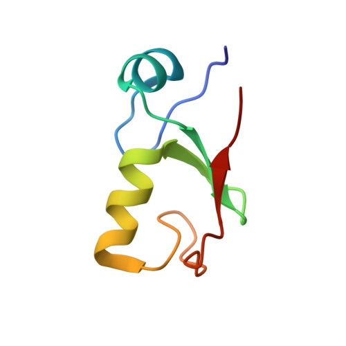
    2XEU

  • PubMed Abstract: 

    Mammalian RNF4 is a dimeric RING ubiquitin E3 ligase that ubiquitylates poly-SUMOylated proteins. We found that RNF4 bound ubiquitin-charged UbcH5a tightly but free UbcH5a weakly. To provide insight into the mechanism of RING-mediated ubiquitylation, we docked the UbcH5~ubiquitin thioester onto the RNF4 RING structure. This revealed that with E2 bound to one monomer of RNF4, the thioester-linked ubiquitin could reach across the dimer to engage the other monomer. In this model, the 'Ile44 hydrophobic patch' of ubiquitin is predicted to engage a conserved tyrosine located at the dimer interface of the RING, and mutation of these residues blocked ubiquitylation activity. Thus, dimeric RING ligases are not simply inert scaffolds that bring substrate and E2-loaded ubiquitin into close proximity. Instead, they facilitate ubiquitin transfer by preferentially binding the E2~ubiquitin thioester across the dimer and activating the thioester bond for catalysis.


  • Organizational Affiliation

    Wellcome Trust Centre for Gene Regulation and Expression, College of Life Sciences, University of Dundee, Dundee, UK.


Macromolecules
Find similar proteins by:  (by identity cutoff)  |  3D Structure
Entity ID: 1
MoleculeChains Sequence LengthOrganismDetailsImage
RING FINGER PROTEIN 464Homo sapiensMutation(s): 0 
UniProt & NIH Common Fund Data Resources
Find proteins for P78317 (Homo sapiens)
Explore P78317 
Go to UniProtKB:  P78317
PHAROS:  P78317
GTEx:  ENSG00000063978 
Entity Groups  
Sequence Clusters30% Identity50% Identity70% Identity90% Identity95% Identity100% Identity
UniProt GroupP78317
Sequence Annotations
Expand
  • Reference Sequence
Oligosaccharides

Help

Entity ID: 2
MoleculeChains Length2D Diagram Glycosylation3D Interactions
beta-D-fructofuranose-(2-1)-alpha-D-glucopyranose
B
2N/A
Glycosylation Resources
GlyTouCan:  G05551OP
GlyCosmos:  G05551OP
Biologically Interesting Molecules (External Reference) 1 Unique
Experimental Data & Validation

Experimental Data

  • Method: X-RAY DIFFRACTION
  • Resolution: 1.50 Å
  • R-Value Free: 0.197 
  • R-Value Work: 0.168 
  • R-Value Observed: 0.170 
  • Space Group: P 41 3 2
Unit Cell:
Length ( Å )Angle ( ˚ )
a = 72.616α = 90
b = 72.616β = 90
c = 72.616γ = 90
Software Package:
Software NamePurpose
REFMACrefinement
HKL-2000data reduction
HKL-2000data scaling
SHELXphasing

Structure Validation

View Full Validation Report


Entry History 

Deposition Data

Revision History  (Full details and data files)

  • Version 1.0: 2010-07-28
    Type: Initial release
  • Version 1.1: 2011-08-24
    Changes: Database references, Other, Refinement description, Version format compliance
  • Version 1.2: 2011-09-14
    Changes: Database references, Other, Refinement description
  • Version 1.3: 2012-10-03
    Changes: Database references
  • Version 2.0: 2020-07-29
    Type: Remediation
    Reason: Carbohydrate remediation
    Changes: Atomic model, Data collection, Derived calculations, Non-polymer description, Other, Structure summary