2XDM

Crystal structure of a complex between Actinomadura R39 DD peptidase and a peptidoglycan mimetic boronate inhibitor


Experimental Data Snapshot

  • Method: X-RAY DIFFRACTION
  • Resolution: 2.40 Å
  • R-Value Free: 0.262 
  • R-Value Work: 0.199 
  • R-Value Observed: 0.203 

wwPDB Validation   3D Report Full Report


Ligand Structure Quality Assessment 


This is version 1.2 of the entry. See complete history


Literature

Crystal Structure of a Complex between the Actinomadura R39 Dd-Peptidase and a Peptidoglycan- Mimetic Boronate Inhibitor: Interpretation of a Transition State Analogue in Terms of Catalytic Mechanism.

Dzhekieva, L.Rocaboy, M.Kerff, F.Charlier, P.Sauvage, E.Pratt, R.F.

(2010) Biochemistry 49: 6411

  • DOI: https://doi.org/10.1021/bi100757c
  • Primary Citation of Related Structures:  
    2XDM

  • PubMed Abstract: 

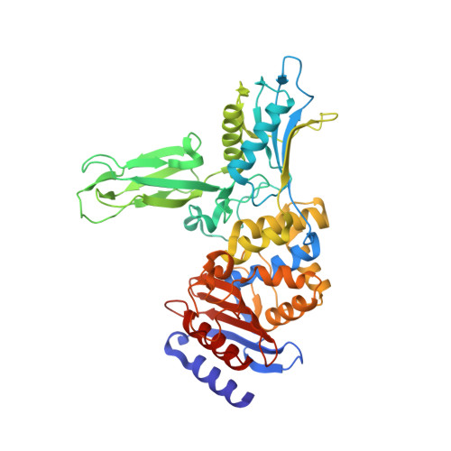
    The Actinomadura R39 DD-peptidase is a bacterial low molecular weight class C penicillin-binding protein. It has previously been shown to catalyze hydrolysis and aminolysis of small D-alanyl-D-alanine terminating peptides, especially those with a side chain that mimics the amino terminus of the stem peptide precursor to the bacterial cell wall. This paper describes the synthesis of (D-alpha-aminopimelylamino)-D-1-ethylboronic acid, designed to be a peptidoglycan-mimetic transition state analogue inhibitor of the R39 DD-peptidase. The boronate was found to be a potent inhibitor of the peptidase with a K(i) value of 32 +/- 6 nM. Since it binds some 30 times more strongly than the analogous peptide substrate, the boronate may well be a transition state analogue. A crystal structure of the inhibitory complex shows the boronate covalently bound to the nucleophilic active site Ser 49. The aminopimelyl side chain is bound into the site previously identified as specific for this moiety. One boronate oxygen is held in the oxyanion hole; the other, occupying the leaving group site of acylation or the nucleophile site of deacylation, appears to be hydrogen-bonded to the hydroxyl group of Ser 298. The Ser 49 oxygen appears to be hydrogen bonded to Lys 52. If it is assumed that this structure does resemble a high-energy tetrahedral intermediate in catalysis, it seems likely that Ser 298 participates as part of a proton transfer chain initiated by Lys 52 or Lys 410 as the primary proton donor/acceptor. The structure, therefore, supports a particular class of mechanism that employs this proton transfer device.


  • Organizational Affiliation

    Department of Chemistry, Wesleyan University, Lawn Avenue, Middletown, CT 06459, USA.


Macromolecules
Find similar proteins by:  (by identity cutoff)  |  3D Structure
Entity ID: 1
MoleculeChains Sequence LengthOrganismDetailsImage
D-ALANYL-D-ALANINE CARBOXYPEPTIDASE
A, B, C, D
466Actinomadura sp. R39Mutation(s): 0 
EC: 3.4.16.4
UniProt
Find proteins for P39045 (Actinomadura sp. (strain R39))
Explore P39045 
Go to UniProtKB:  P39045
Entity Groups  
Sequence Clusters30% Identity50% Identity70% Identity90% Identity95% Identity100% Identity
UniProt GroupP39045
Sequence Annotations
Expand
  • Reference Sequence
Small Molecules
Ligands 4 Unique
IDChains Name / Formula / InChI Key2D Diagram3D Interactions
BO8
Query on BO8

Download Ideal Coordinates CCD File 
L [auth B],
R [auth C]
(D-ALPHA-AMINOPIMELYLAMINO)-D-1-ETHYLBORONIC ACID
C9 H20 B N2 O6
ZSMKWHGOAVMEJJ-RNFRBKRXSA-N
MES
Query on MES

Download Ideal Coordinates CCD File 
EA [auth D]2-(N-MORPHOLINO)-ETHANESULFONIC ACID
C6 H13 N O4 S
SXGZJKUKBWWHRA-UHFFFAOYSA-N
SO4
Query on SO4

Download Ideal Coordinates CCD File 
AA [auth D]
BA [auth D]
E [auth A]
F [auth A]
G [auth A]
AA [auth D],
BA [auth D],
E [auth A],
F [auth A],
G [auth A],
H [auth A],
I [auth A],
M [auth B],
N [auth B],
O [auth B],
P [auth B],
Q [auth B],
S [auth C],
T [auth C],
U [auth C],
V [auth C],
W [auth C],
X [auth D],
Y [auth D],
Z [auth D]
SULFATE ION
O4 S
QAOWNCQODCNURD-UHFFFAOYSA-L
CO
Query on CO

Download Ideal Coordinates CCD File 
CA [auth D],
DA [auth D],
J [auth A],
K [auth A]
COBALT (II) ION
Co
XLJKHNWPARRRJB-UHFFFAOYSA-N
Binding Affinity Annotations 
IDSourceBinding Affinity
BO8 PDBBind:  2XDM Ki: 32 (nM) from 1 assay(s)
Experimental Data & Validation

Experimental Data

  • Method: X-RAY DIFFRACTION
  • Resolution: 2.40 Å
  • R-Value Free: 0.262 
  • R-Value Work: 0.199 
  • R-Value Observed: 0.203 
  • Space Group: P 1 21 1
Unit Cell:
Length ( Å )Angle ( ˚ )
a = 103.308α = 90
b = 91.568β = 94.37
c = 106.871γ = 90
Software Package:
Software NamePurpose
REFMACrefinement

Structure Validation

View Full Validation Report


Ligand Structure Quality Assessment 


Entry History 

Deposition Data

Revision History  (Full details and data files)

  • Version 1.0: 2010-07-21
    Type: Initial release
  • Version 1.1: 2011-11-23
    Changes: Database references, Refinement description, Version format compliance
  • Version 1.2: 2023-12-20
    Changes: Data collection, Database references, Derived calculations, Other, Refinement description